
Przodkiem mikroskopii sił atomowych jest Skaningowy Mikroskop Tunelowy (STM), który został pierwotnie opracowany w 1981 roku przez dwóch naukowców pracujących w IBM w Szwajcarii, Gerda Binniga i Heinricha Rohrera. Głównym celem było uzyskanie topograficznego obrazu powierzchni próbki z wykorzystaniem sił oddziaływań międzyatomowych. W 1986 roku otrzymali oni za ten wynalazek Nagrodę Nobla w dziedzinie fizyki. Od tego czasu rodzina mikroskopów z sondą skanującą wciąż się rozwija. [1] Obecnie jesteśmy w stanie obserwować pojedyncze atomy i mierzyć siły, które pomiędzy nimi występują.
The ancestor of atomic force microscopy is Scanning Tunneling Microscope (STM), which was originally developed in 1981 by two scientists while working at IBM in Switzerland, Gerd Binnig and Heinrich Rohrer. The main idea was to obtain a topographic image of the sample surface using the forces of the interatomic interactions. In 1986 they were awarded the Nobel Prize in physics for this invention. Since that time, the family of scanning probe microscopes is still developing.[1] We are now able to observe single atoms and measure the forces that occurs between them.
Używając STM możemy obrazować tylko próbki, które są przewodzące lub półprzewodzące, jednak w 1986 roku Gerd Binnig, Calvin F. Quate i Christoph Gerber zbudowali pierwszy Mikroskop Sił Atomowych AFM, aby przezwyciężyć to ograniczenie. [2] Używając AFM możemy zmierzyć prawie każdy rodzaj powierzchni, nawet próbki biologiczne!
Using STM we can image only samples which are conducting or semiconducting, but in 1986 Gerd Binnig, Calvin F. Quate and Christoph Gerber built the first AFM to overcome this limitation. [2] Using AFM we can measure almost every type of the surface, even biological samples!
Ale jak on działa?/But how it works?
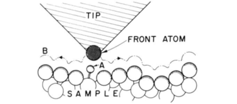
Zbieramy informacje o mierzonej próbce na podstawie interakcji pomiędzy ostrą końcówką tak zwanego tipa a powierzchnią próbki. Gdy mechaniczna sonda porusza się po próbce i dotyka jej lub utrzymuje stałą, bardzo bliską odległość względem jej powierzchni, możemy zebrać informacje o siłach atomowych, które występują pomiędzy tipem a atomami na powierzchni próbki i stworzyć jej obraz topograficzny. [1,3] Idealny tip powinien być zakończony pojedynczym atomem (Rysunek 1), jednak w rzeczywistości jest mniej idealny.
We get the information from the interaction between sharp tip and the surface of measured sample. As the mechanical probe is moving across the sample and touching it or keeping the constant, very close distance from it, we can gather the information about atomic forces that are occurring between the tip and atoms on the surface and create the topographic image. [1,3] The ideal tip should be ended by a single atom (Figure 1), however in reality it is less perfect.
Ostro zakończony tip jest przymocowany do wspornika umieszczonego nad próbką Wspornik ten odchyla się pod wpływem sił działających pomiędzy tipem a mierzoną powierzchnią (rys. 2a). Gdy tip zbliża się do powierzchni, siły przyciągające, które występują w bliskim zasięgu ze względu na siły van der Waalsa, odchylają ją w kierunku powierzchni. Jednak, kiedy tip niemal dotyka powierzchni, siły odpychające wzrastają i odchylają wspornik od powierzchni próbki. Na rysunku 2b widzimy wykres sił działających na tipa w zależności od jego odległości od próbki. [5,6]
The sharp tip is attached to the cantilever placed over the sample, that is deflecting upon the forces acting between the tip and measured surface (Figure 2a). As the tip approaches the surface, the attractive forces, that are occurring in close-range due to van der Waals force, are deflecting it to the surface. But when the tip is almost touching the surface, the repulsive forces are increasing and deflecting the cantilever away. On the Figure 2b, we can see a graph of the forces acting on the tip, depending on the distance from the sample. [5,6]
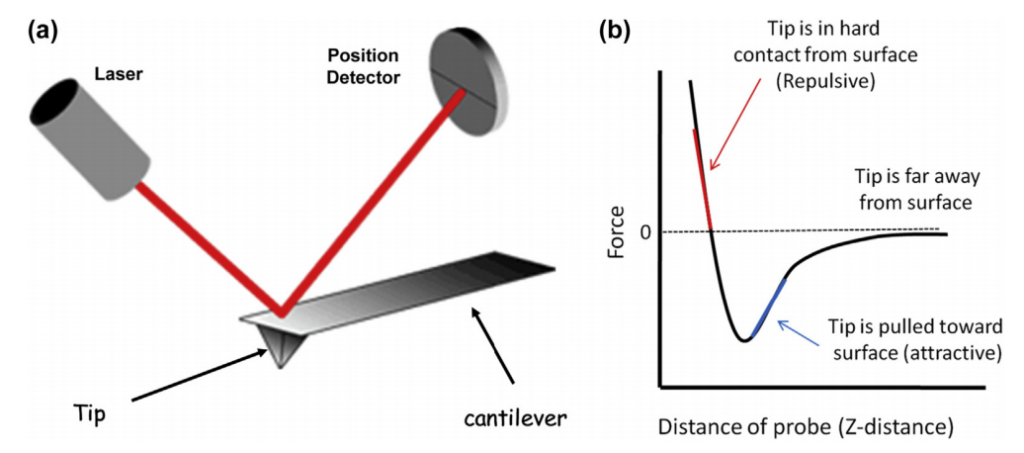
Do wykrywania tych ugięć wspornika wykorzystywana jest wiązka laserowa (rys 2a). Kąt wiązki odbitej od górnej części wspornika zmienia się nieznacznie w zależności od przemieszczenia wspornika. Informacja o zmianie położenia wiązki laserowej jest zbierana przez czułą fotodiodę i dalej przetwarzana na sygnał elektryczny, który ostatecznie jest przekształcany na kolorową mapę wyrażającą topografię próbki w postaci odcieni gamy kolorów. [1,6]
To detect those deflections a laser beam is used (Figure 2a). The angle of beam reflected from the top of cantilever is slightly changing depending on the cantilever displacement. This change in laser beam position is collected by position sensitive photodiode and further converted into electrical signal, that is finally converted into colored map expressing the sample topography in a form of hues. [1,6]
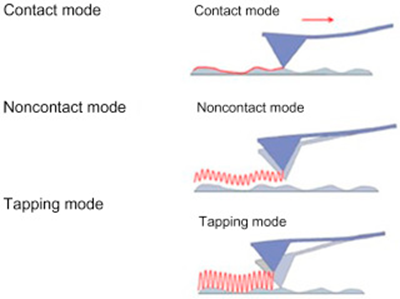
Istnieją 3 tryby obrazowania AFM, które są zależne od typu mierzonej próbki.
– Tryb kontaktowy – w którym końcówka stale dotyka powierzchni i jest „ciągnięta” po próbce, dlatego nie może być zbyt sztywna, aby się nie złamała. Siły działające na wspornik są prawie zawsze siłami odpychającymi. Ten tryb jest zwykle stosowany do pomiarów twardych powierzchni.
– Tryb kontaktu przerywanego – to dynamiczny tryb kontaktu. Może być stosowany w przypadku delikatniejszych próbek i płynów. Tutaj wspornik bardzo szybko oscyluje w górę iw dół, blisko częstotliwości rezonansowej. Tip pozostaje w zasięgu sił bliskiego zasięgu, ale nie ma ryzyka, że przyklei się do próbki. Jest to najczęściej stosowany tryb pracy AFM.
– Tryb bezkontaktowy – w którym końcówka oscyluje nad próbką i nigdy jej nie dotyka. W tym trybie wykorzystywane są siły dalekiego zasięgu, takie jak siły magnetyczne, elektrostatyczne lub przyciągające siły van der Waalsa. Mierzone są zmiany częstotliwości rezonansowej. [2,3]
There are 3 modes of the AFM imaging, depend on the measured sample.
– Contact mode – in which, the tip is constantly touching the surface and is “dragged” across the sample, thus it can’t be too stiff so it doesn’t break. Forces acting on the cantilever are almost always repulsive forces, and this mode is usually done with solid surfaces.
– Tapping mode – is a dynamic contact mode. It can be used with more delicate samples and with liquids. Here, the cantilever is oscillating very quickly up and down, near to its resonance frequency. The tip is staying in the range of short-range forces, but there is no risk, that it would stick to the sample. It is the most commonly used mode.
– Non-contact mode – in which the tip is oscillating above the sample and never touching it. In this mode long-range forces are used, like magnetic, electrostatic, or attractive van der Waals forces and the changes in the resonance frequency are measured. [2,3]
Dzięki mikroskopowi sił atomowych możemy uzyskać topografię powierzchni mierzonej próbki, ale także jej właściwości fizyczne, takie jak: tarcie, przyczepność, rozkład ładunków elektrostatycznych, przewodnictwo elektryczne i wiele innych. Przeprowadzenie pomiaru zwykle nie wymaga skomplikowanych procedur przygotowania badanej próbki i może być dokonane zarówno w powietrzu, jak i w cieczy czy w próżni, dzięki czemu możliwe jest badanie właściwości żywych komórek w ich naturalnym ciekłym środowisku. Pośród innych licznych zalet AFM jest także możliwość wygenerowania struktury 3D mierzonej powierzchni na podstawie zebranych danych. Oczywiście są ta technika ma również swoje ograniczenia. Jedną z wad jest szybkość skanowania – pomiar pojedynczego skanowania zajmuje kilka minut, co może skutkować zniekształceniami obrazu. Istnieje również ryzyko występowania artefaktów na obrazie, a rozmiar pojedynczego skanu jest ograniczony. Możliwości zastosowania AFM jednak wciąż rosną. Można go łączyć z innymi technikami mikroskopii optycznej i spektroskopii, co daje początek wielu nowym metodom pomiarowym, np. skaningowej mikroskopii optycznej pola bliskiego albo nano-FTIR. [2,3]
Using AFM we can obtain the surface topography of measured sample, but also its physical properties such as: friction, adhesion, electrostatic charge distribution, electrical conductivity and more. The measurement usually does not require complicated sample preparation procedures and can be performed both in air, in a liquid or in a vacuum, making it possible to study the properties of living cells in their natural liquid environment. Among other numerous advantages of the AFM is the ability to generate the 3D structure of measured surface from collected data. Of course, there are some limitations of this technique too. One of the disadvantages is the scanning speed – single scan takes few minutes to be measured, what can result in some image distortions. There is also possibility of image artefacts and single scan image size is limited. However, the applicability of AFM continues to grow It can be also combined with other optical microscopy and spectroscopy techniques, giving rise to many new measurement methods e.g., scanning near-field optical microscopy and nano-FTIR. [2,3]
Reasumując, AFM jest niesamowitą techniką wykorzystującą siły oddziaływań międzyatomowych, dzięki której jesteśmy w stanie zmierzyć niemal każdy rodzaj materiału o zdolności rozdzielczej rzędu wymiarów pojedynczego atomu!
AFM is amazing technique using the forces of interatomic interactions, thanks to which we are able to measure almost every type of material with the resolving power of the order of dimensions of a single atom!
Bibliografia/References:
[1] https://www.nanoscience.com/techniques/atomic-force-microscopy/
[2] https://pl.wikipedia.org/wiki/Mikroskop_si%C5%82_atomowych
[3] https://en.wikipedia.org/wiki/Atomic_force_microscopy
[4] G. Binnig, C. F. Quate, Ch. Gerber, Atomic Force Microscope, Physical Review Letters, 1986, 56 (9), 930-933
[5] V. Sudarsan, (2017), ‘Materials for hostile chemical environments’, in A.K. Tyagi and S. Banerjee (ed.) Materials Under Extreme Conditions, p. 135
[6] https://www.parksystems.com/medias/nano-academy/how-afm-works
[7] R. Asmatulu and W. S. Khan, Synthesis and Applications of Electrospun Nanofibers, 2019, p. 266