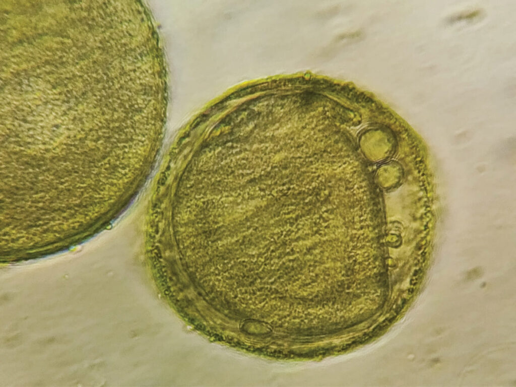
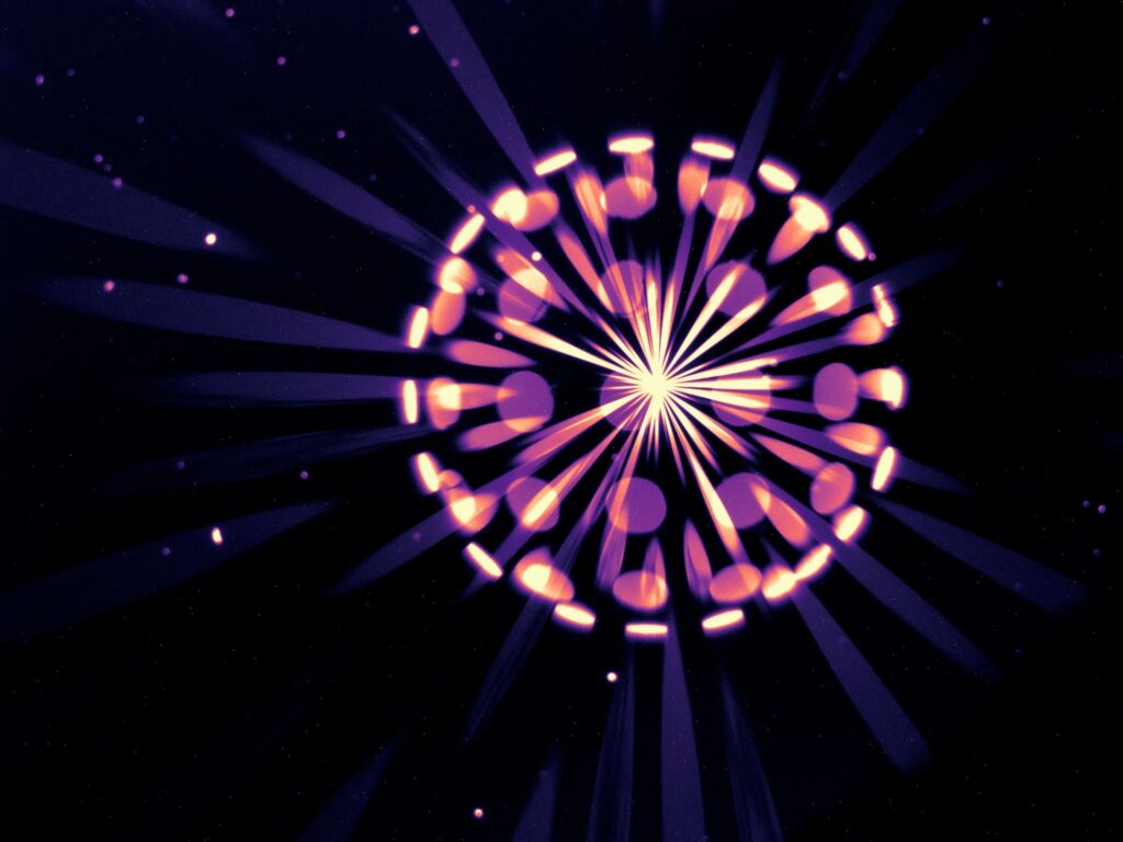
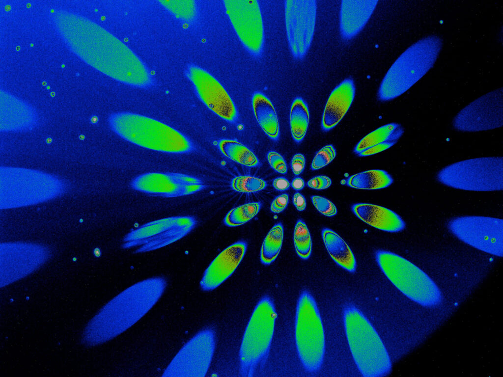
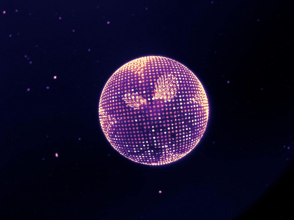
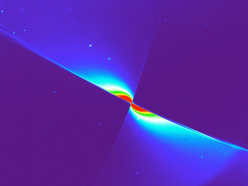
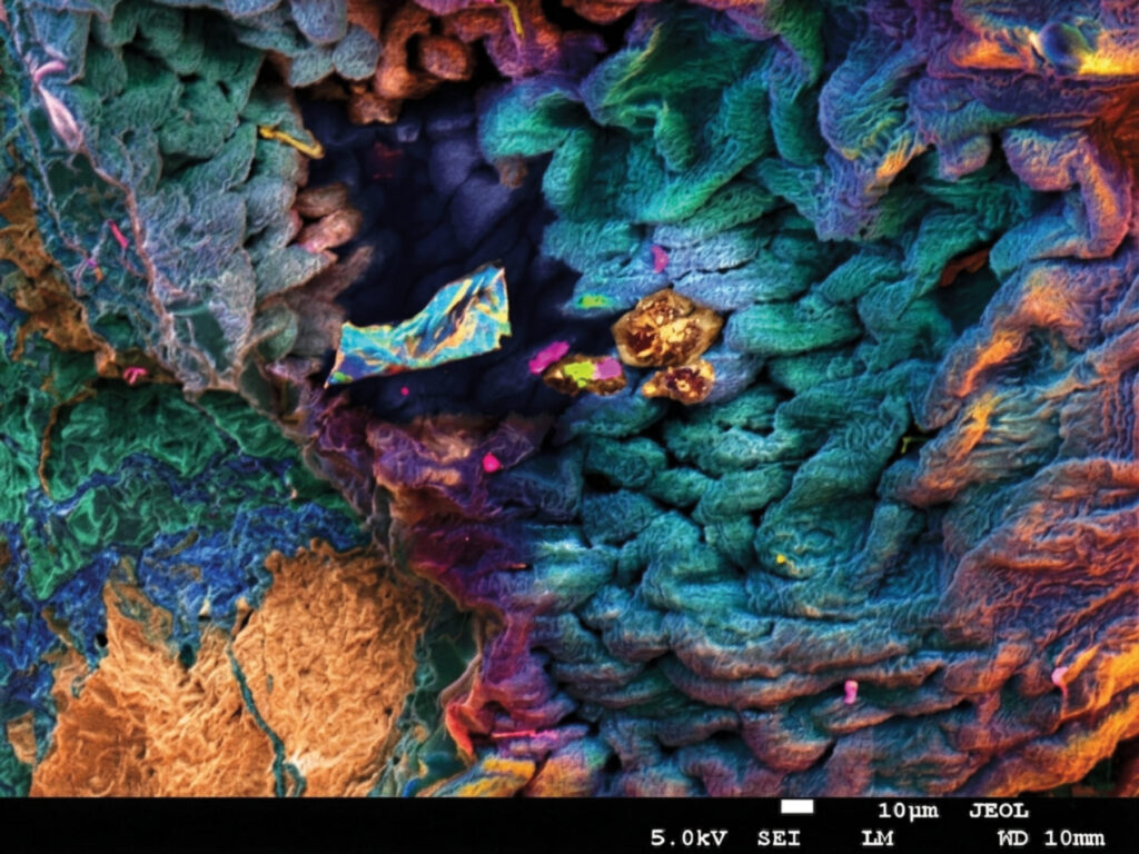
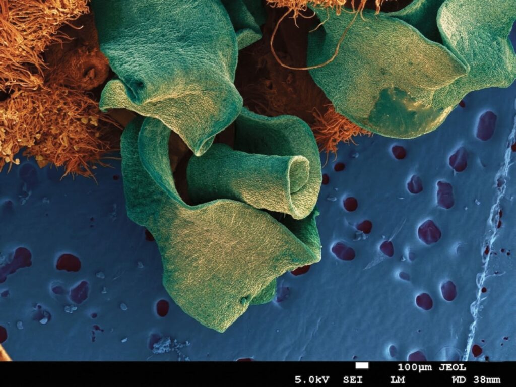
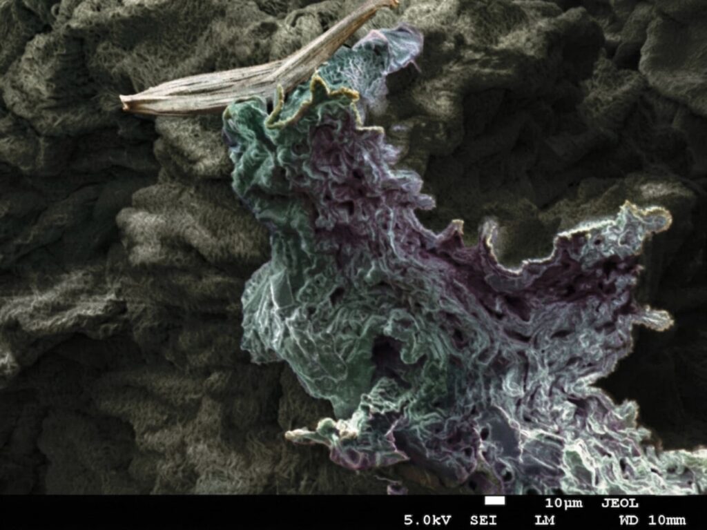
Here, you can find all of the record-breaking 153 photos that took part in this year’s contest.
The winning artworks:
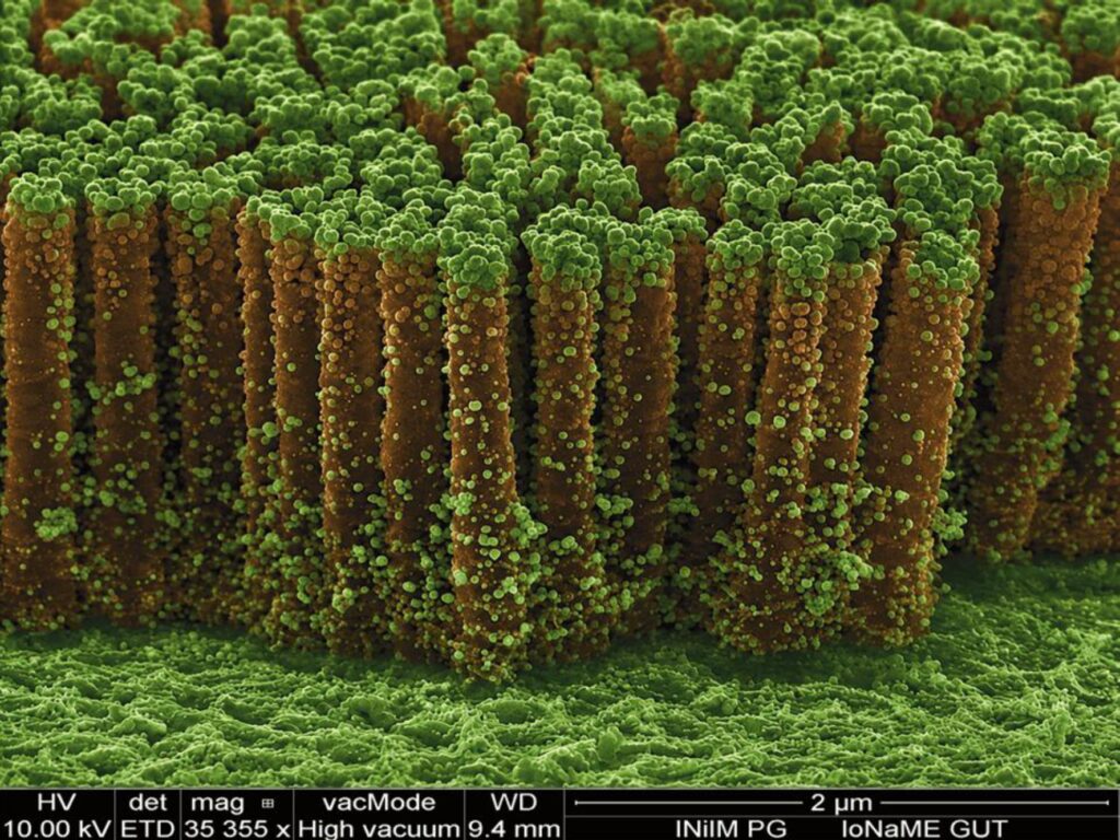
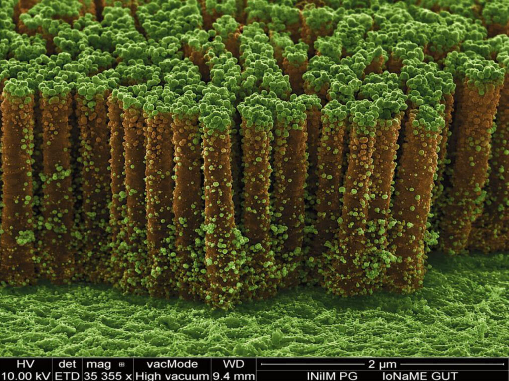
1st place – Nanojungle by Stefania Wolff, Faculty of Applied Physics and Mathematics, Gdańsk University of Technology and Centre of Plasma and Laser Engineering, Institute of Fluid-Flow Machinery, Polish Academy of Sciences
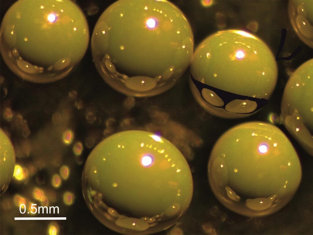
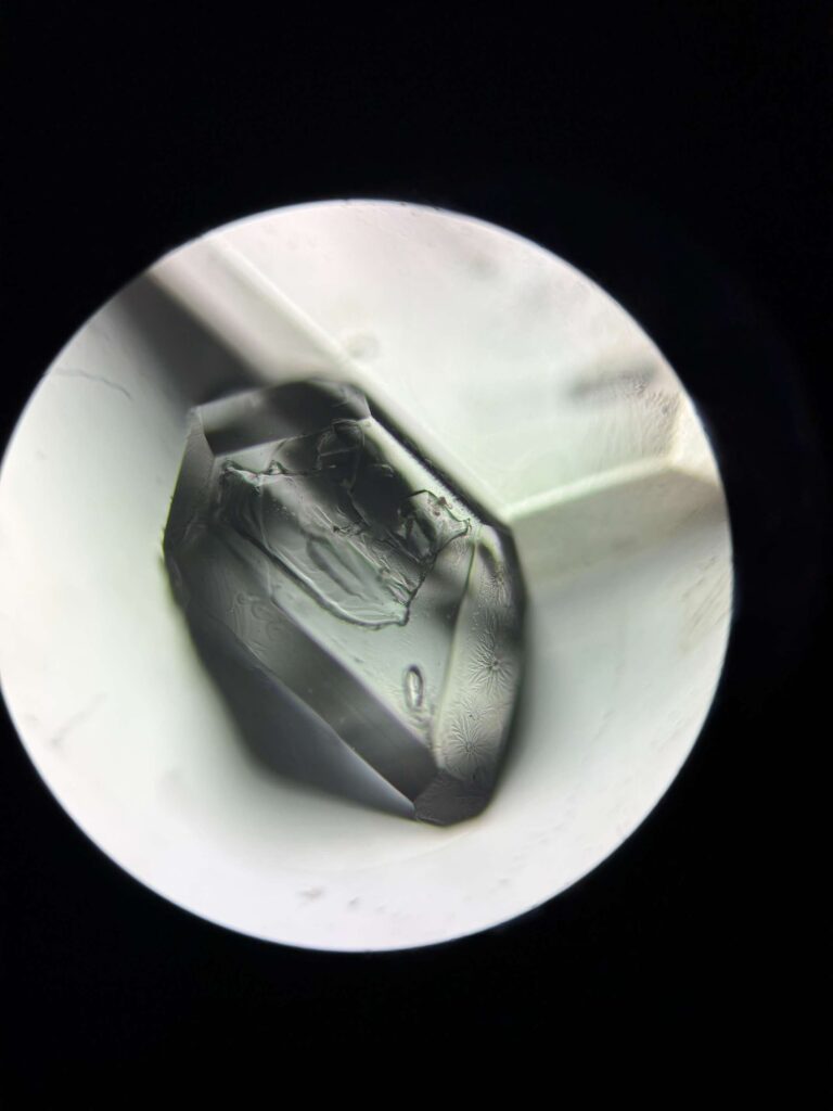
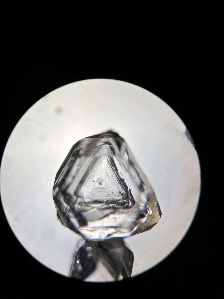
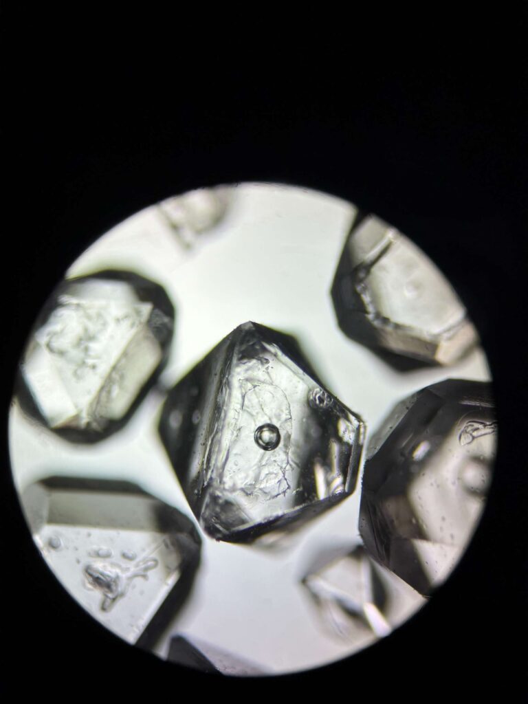
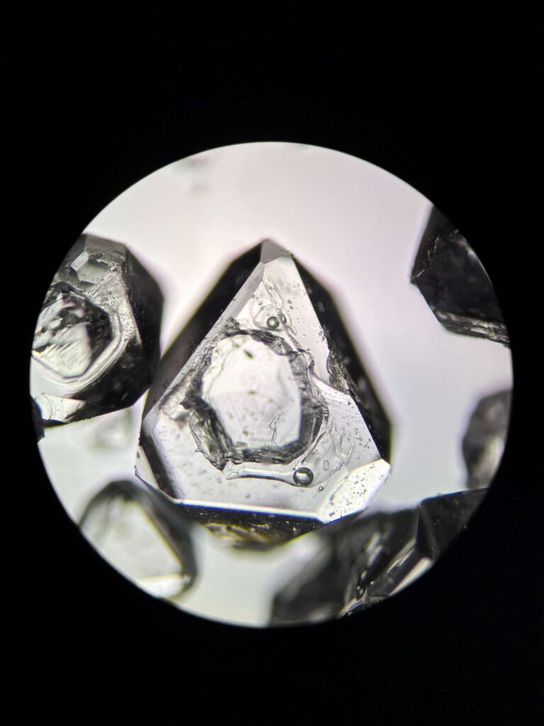
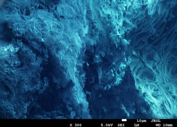
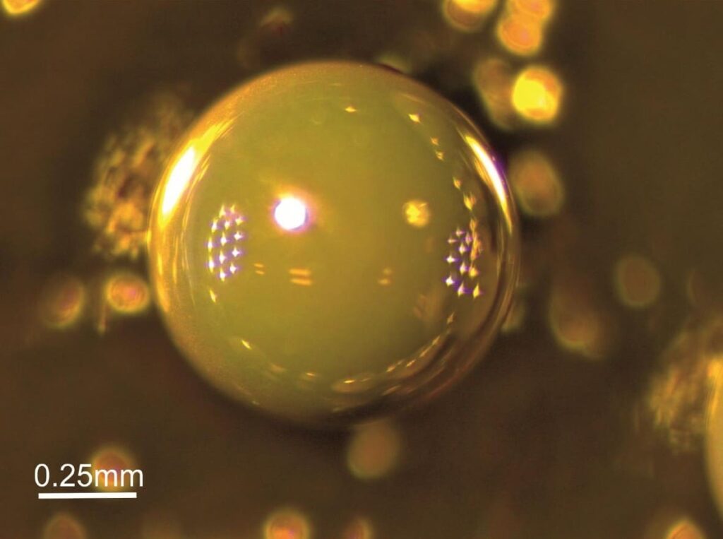
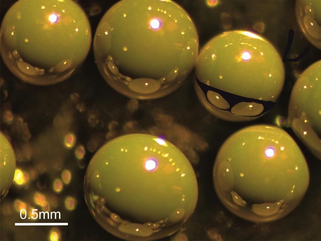
2nd place – micro gangsters attack by Zofia Mazurkiewicz, Institute of Low Temperature and Structure Research, Polish Academy of Sciences
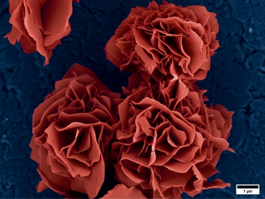
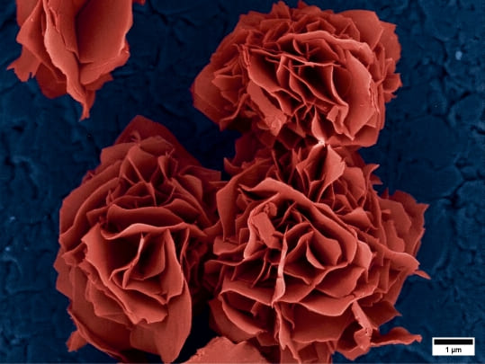
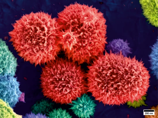
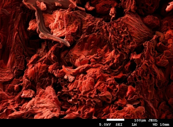
3rd place – The Rose of Ceres by Damian Szymański, Institute of Low Temperature and Structure Research, Polish Academy of Sciences
MANIAC 2026 was financed by ORLEN Foundation.
Below you can find all works sorted alphabetically by Author first name:
Alica Christophoryova – Department of Zoology, Faculty of Natural Sciences, Comenius University
I am a graphic designer and technical specialist at the Department of Zoology, Comenius University Bratislava. My work includes scientific photography and graphic materials for publications focused on invertebrates.
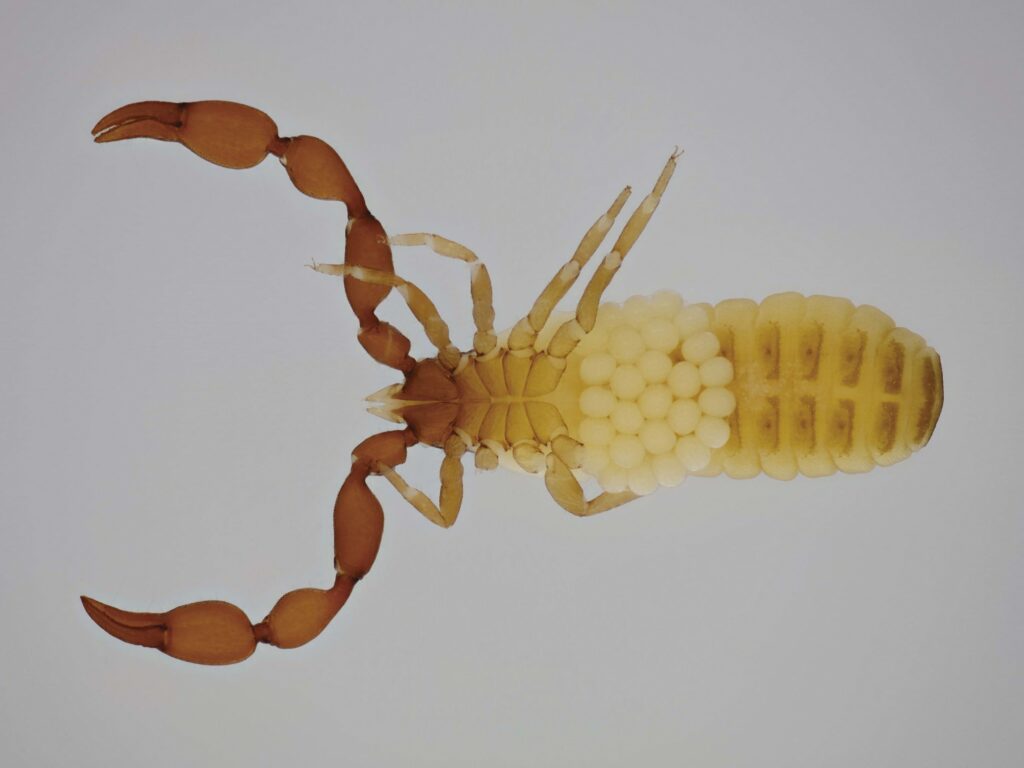
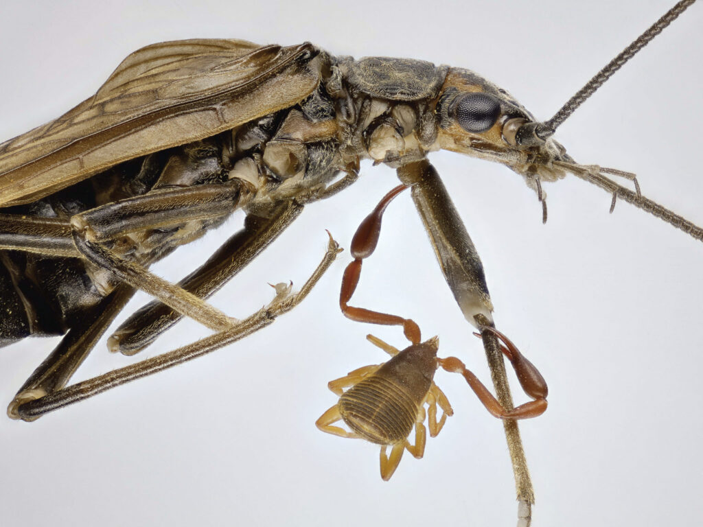
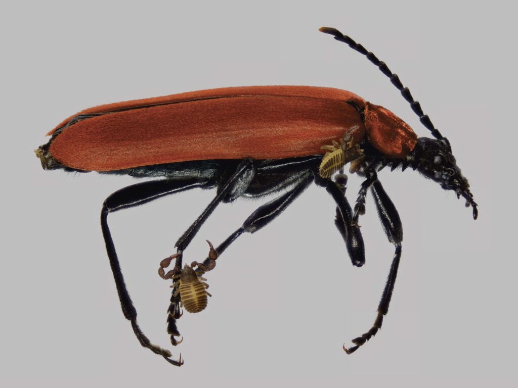
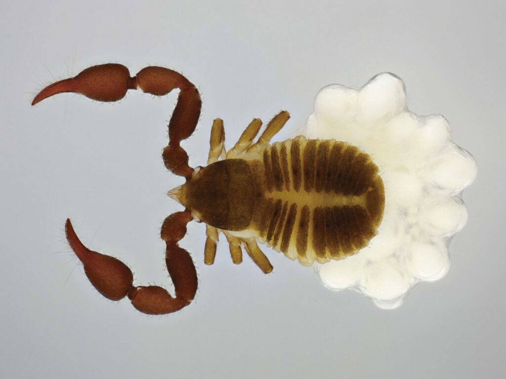
Bearer of Life – Female of the pseudoscorpion Lamprochernes chyzeri carrying a brood sac was taken using a Canon EOS 5D Mark II camera attached to a Zeiss Axio Zoom V16 stereomicroscope.Hitchhiker – Phoresy of the pseudoscorpion Mesochelifer ressli on the stonefly Perlodes microcephalus was taken using a Canon EOS 5D Mark II camera attached to a Zeiss Axio Zoom V16 stereomicroscope.Silent Passengers – Phoresy of the pseudoscorpion Chernes hahnii on the beetle Pyrochroa coccinea was taken using a Canon EOS 5D Mark II camera attached to a Zeiss Axio Zoom V16 stereomicroscope. The Broodkeeper – Female of the pseudoscorpion Chernes hahnii carrying eggs in a brood sac was taken using a Canon EOS 5D Mark II camera attached to a Zeiss Axio Zoom V16 stereomicroscope.
Amelia Maj – Wrocław University of Science and Technology
I am a first-cycle student at the Faculty of Chemistry. My scientific interests include: intersection of chemistry and biology, applications of electron microscopy in biology and medicine, TEM.
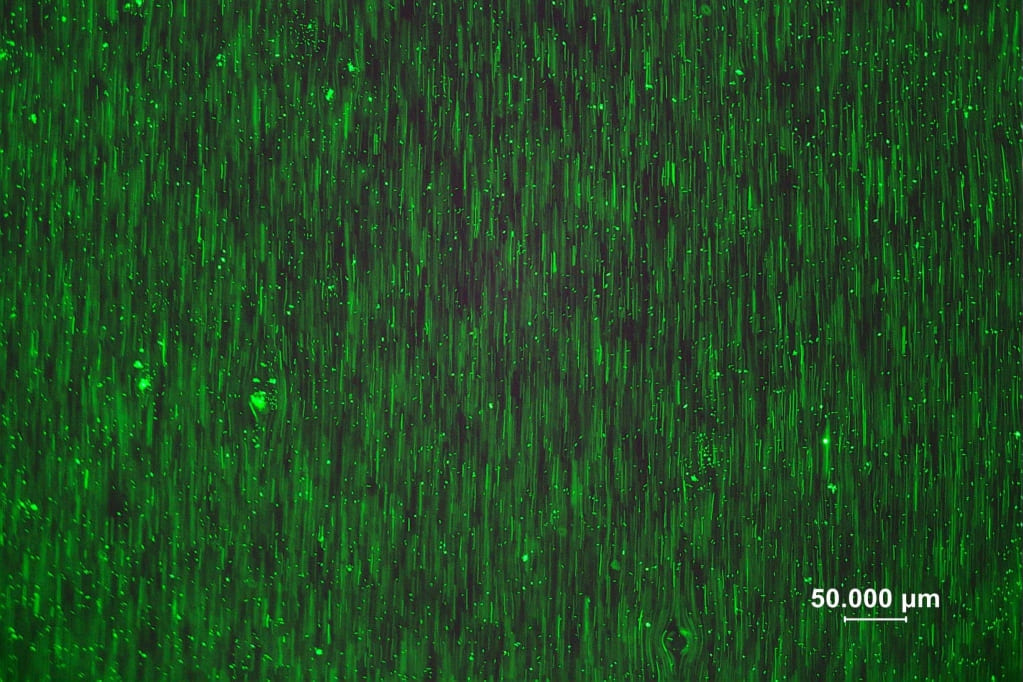
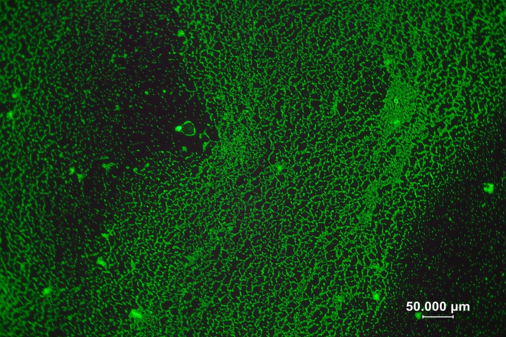
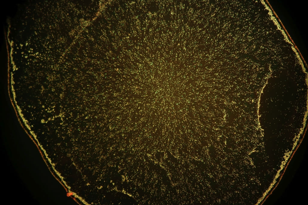
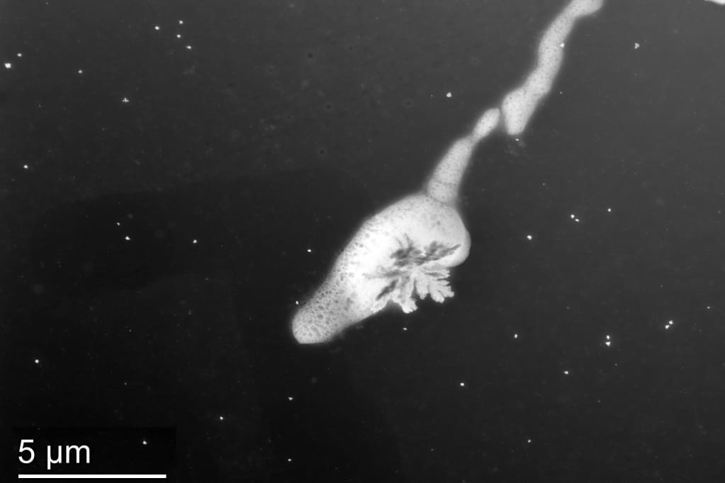
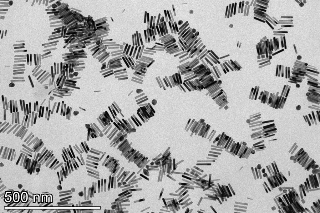
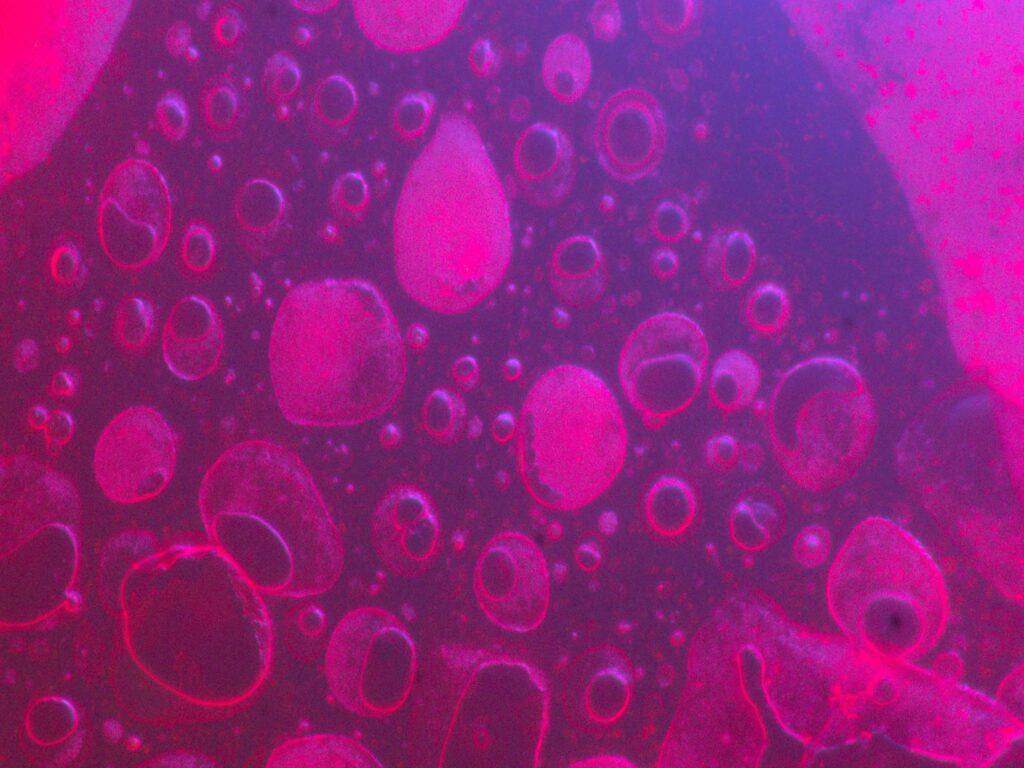
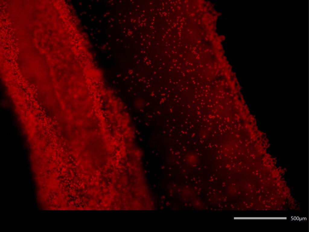
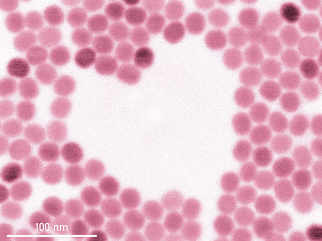
Matrix of Life – Fluorescence microscopy, Acinetobacter baumanii, dyed with LIVE/DEAD kit Fluorescent Reef – Fluorescence microscopy, Acinetobacter baumanii, dyed with LIVE/DEAD kit Microcosmos – Fluorescence microscopy, Acinetobacter baumanii, dyed with LIVE/DEAD kit Bloom in the Void – Scanning transmission electron microscopy, ionic liquid Toward Alignment – Transmission electron microscopy, gold nanoparticles
Anna Hübner – University of Rijeka
4th year medical student, 23 years old
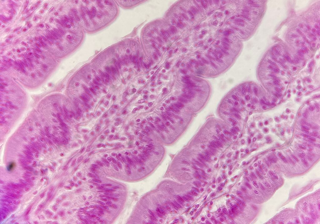
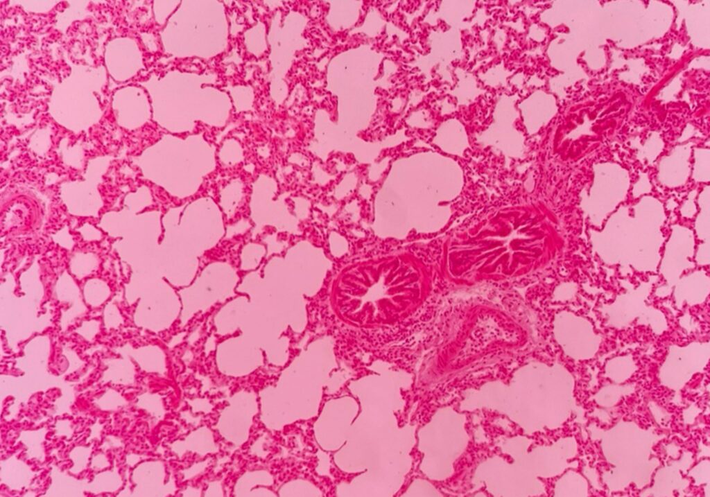
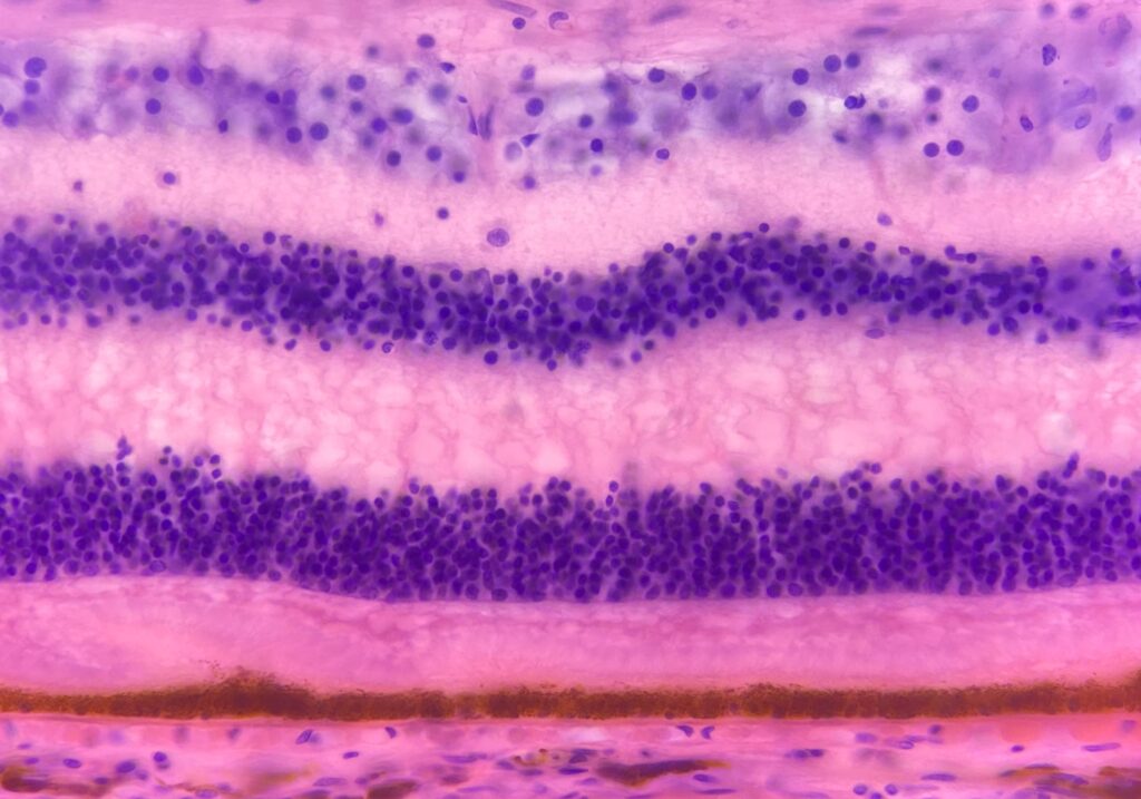
Scientific Hunger – The picture shows intestinal villi photographed by a light microscope. It is a histological slide with H&E staining.Breathe! – The picture shows healthy lung tissue through a light microscope. It is a histological slide in H&E staining. Colors of our rainbow – This is a microscopic photo through a light microscope. It shows the layers of our retina.
Bartłomiej Cichy – Institute of Low Temperature and Structure Research, Polish Academy of Sciences
The author works in the field of quantum photonics and spin-light-matter interactions. His primary focus is on quantum light generation and quantum imaging using single and coupled nanosystems.
10 nm from chaos – High-angle annular dark-field scanning transmission electron microscopy. The artwork is derived from microscopic structures observed during research on nanomaterials. The photo shows an SiO₂ nanoparticle with CdSe quantum dots embedded on its surface.
Bartłomiej Łuszczuk – Faculty of Chemistry, Warsaw University of Technology
A Medical Biotechnology student at Warsaw University of Technology specializing in organ-on-a-chip technology. By merging engineering with biology, he explores the intersection of microfluidics and living systems. His photography brings the hidden aesthetics of the microscopic world to a broader audience.
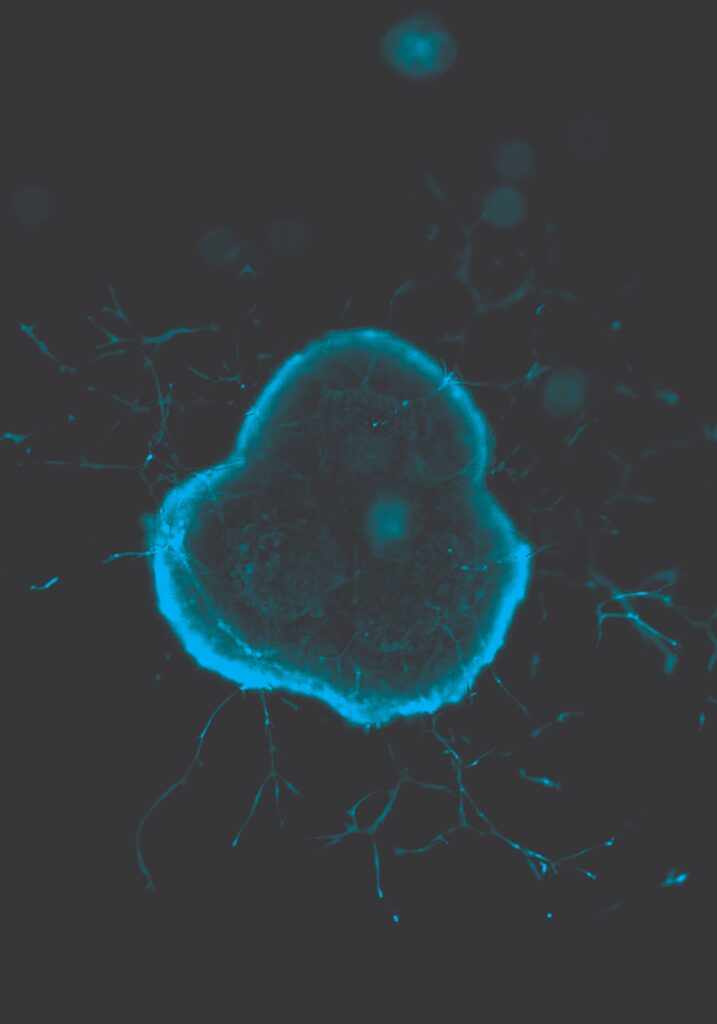
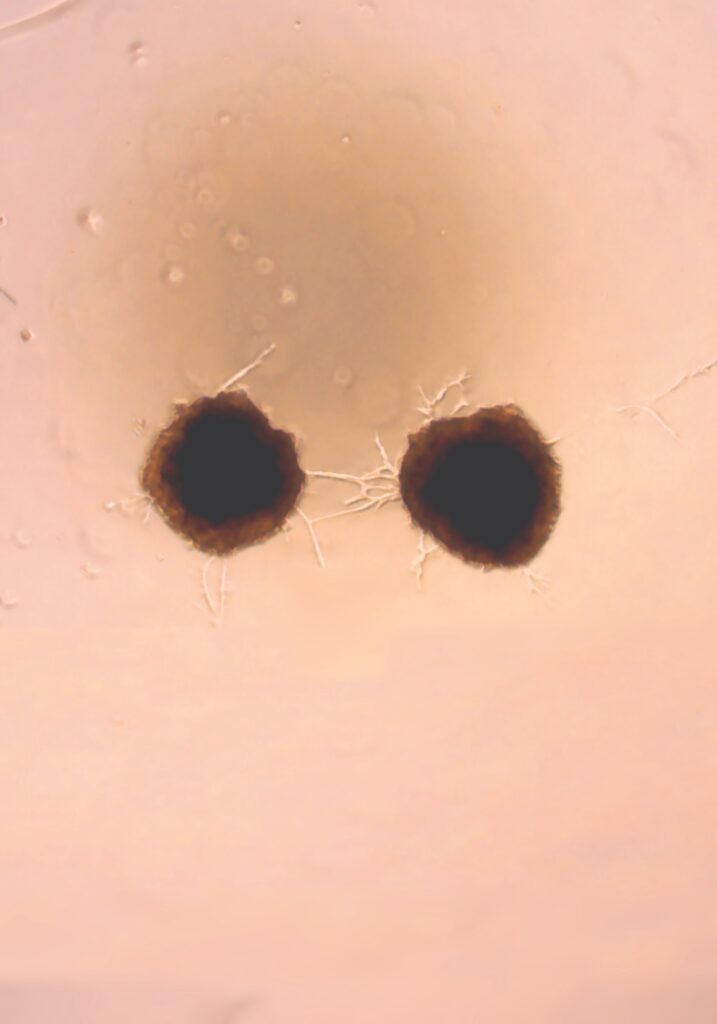
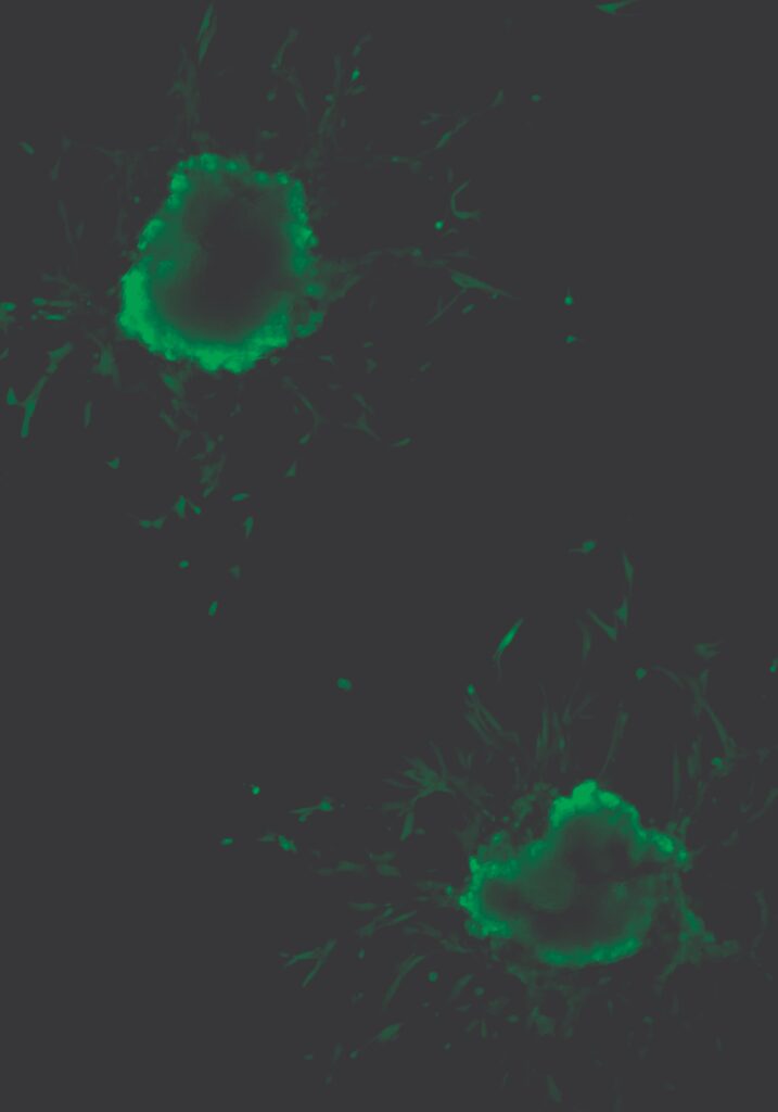
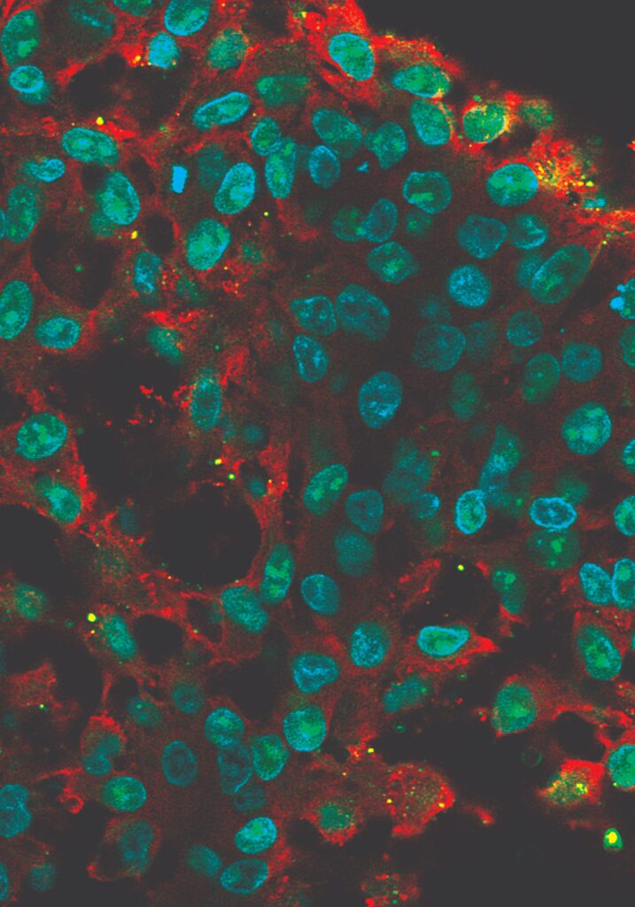
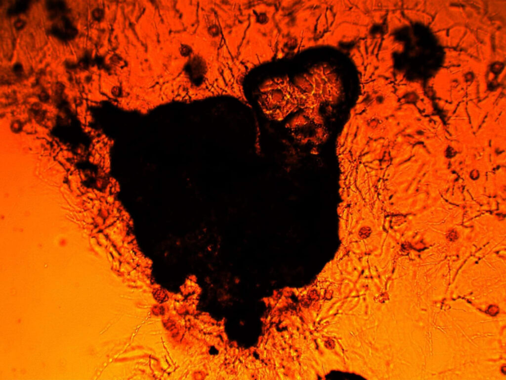
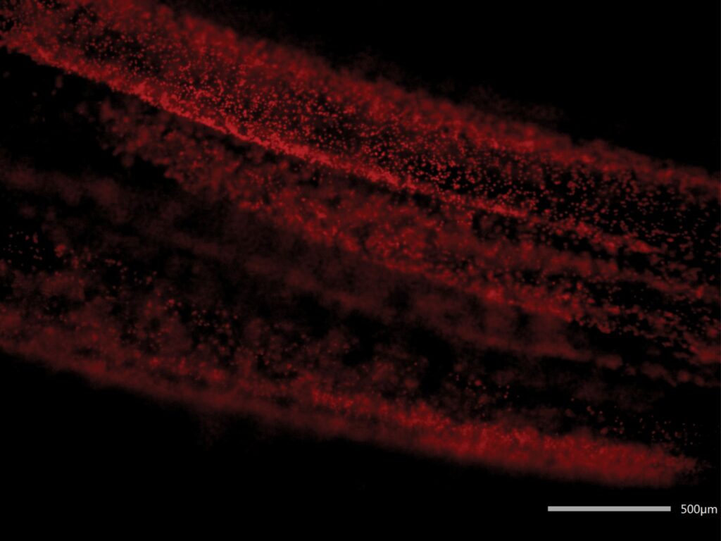
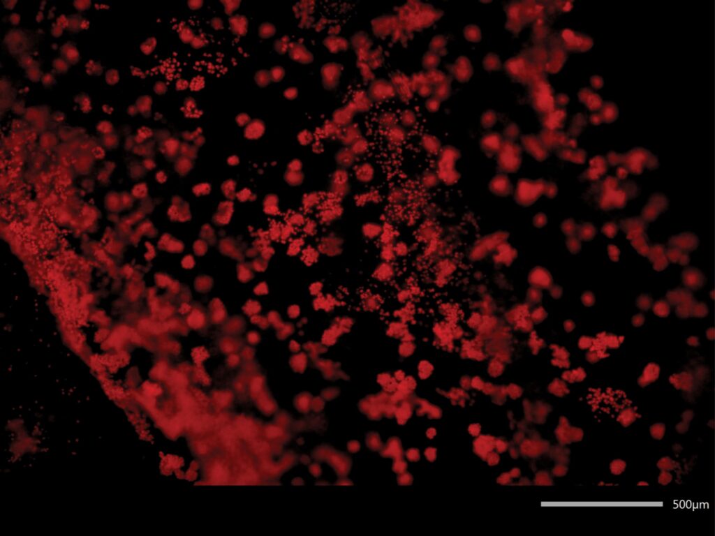
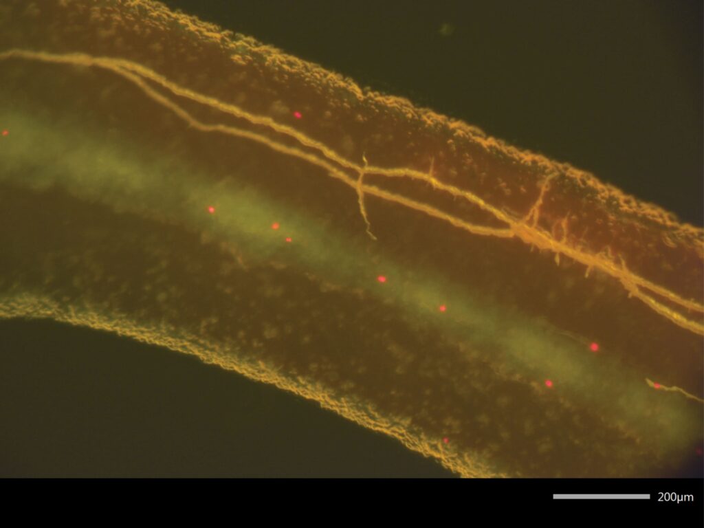
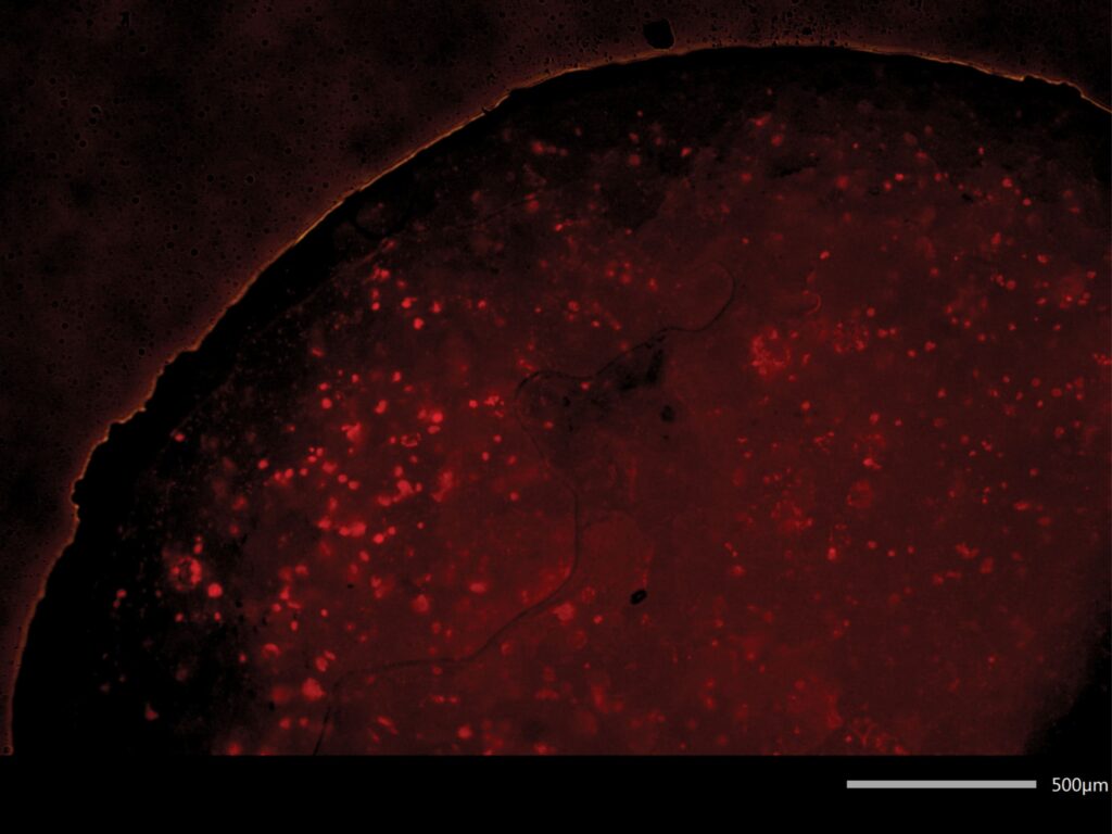
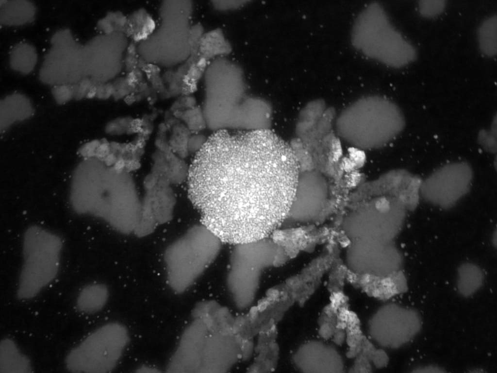
Mind Flyer – Cancer model aggregate in gel (Fluorescence microscopy). Description: A dark monolith of malignancy extending its “demonic” tendrils to colonize the matrix, echoing the haunting anatomy of the Stranger Things namesake. Captured via fluorescence, it reveals a biological shadow-realm, illustrating the relentless expansion of a parasite determined to dominate its environment. The creation of Adam – Cellular aggregates in contact (EVOS digital optical microscopy). Description: A serendipitous reflection of Michelangelo’s masterpiece found within the micro-scale, where two cellular worlds reach out to bridge the void. This accidental encounter captures the precise moment of biological communion, proving that even in the smallest structures, art and life converge.Spheroid Bloom – Twin aggregates in Matrigel (Fluorescence microscopy). Description: Emerging from the darkness of the Matrigel, these twin orbs flourish as they extend pioneer shoots in a desperate search for sustenance. The fluorescent glow documents a silent, expansive dance of survival, where every sprouting filament is a testament to the primal hunger of living tissue. Scaffold of Excess – Immunostained liver fibrosis (Confocal microscopy). Description: The moment architecture becomes oppression: here, the crimson collagen transforms from a supportive framework into a suffocating cage. This confocal study reveals the tragic threshold of pathophysiology, where the very material meant to protect the cells begins to constrict the life it once sustained.
Beatrice Ballo – Babeș-Bolyai University
Hi! I’m Bea and I’m 21. I love to take photos, draw, paint and research. I loved to work for this contest.
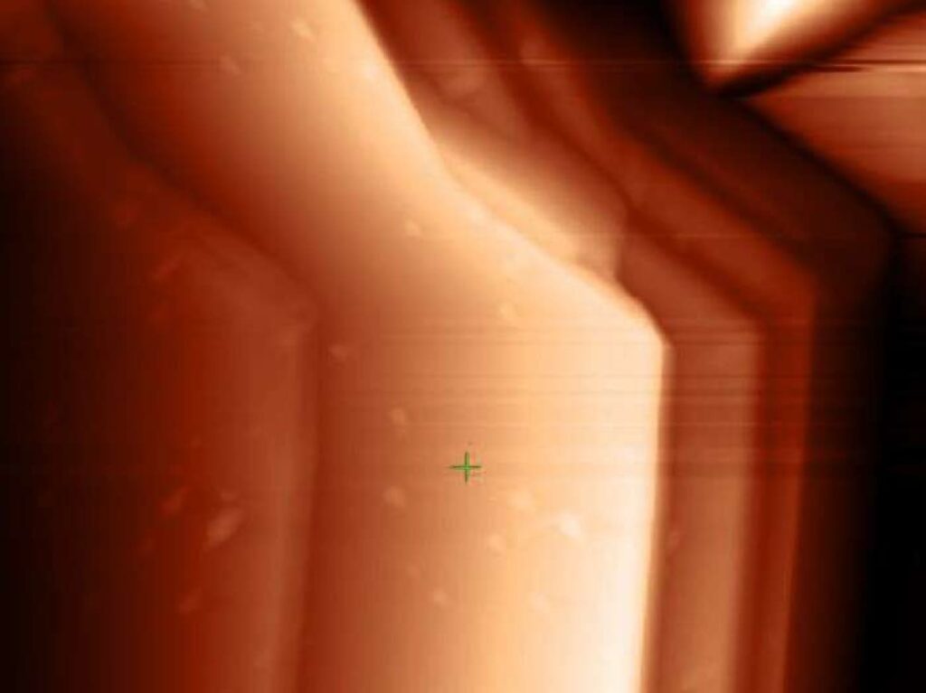
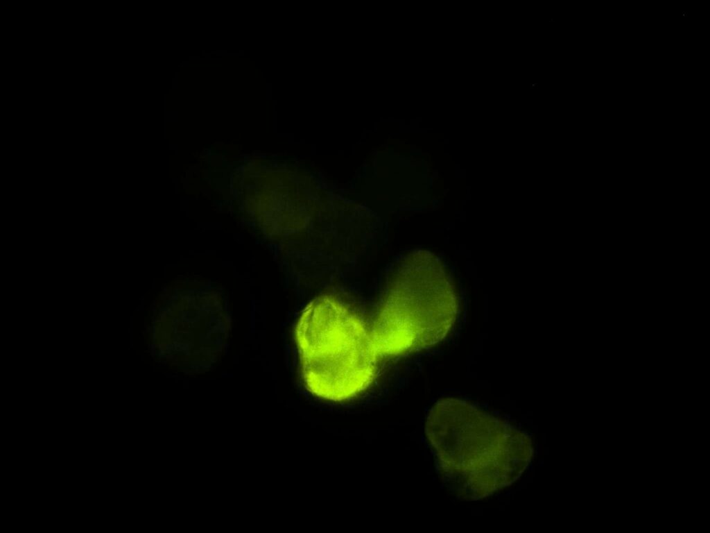
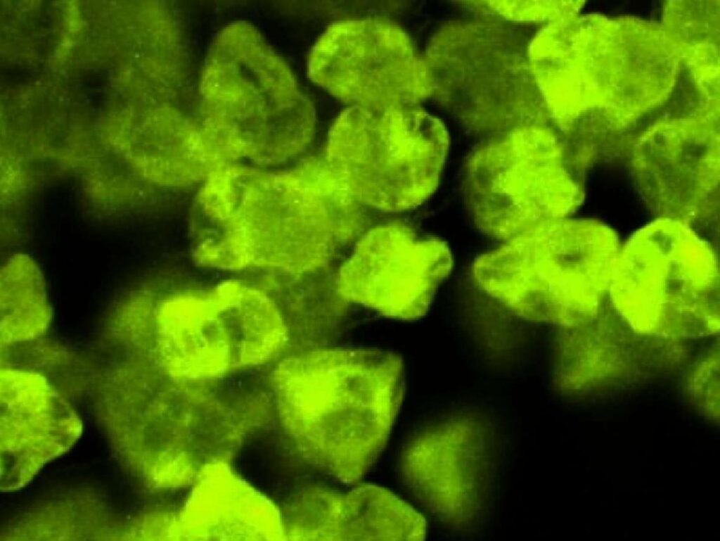
ET – A distorted transmission reveals a glowing extraterrestrial figure emerging through waves of static, its neon outline flickering against the darkness like a mysterious signal from another world. Grand Canyon – This atomic force microscopy image of the Grand Canyon transforms the landscape into a surreal terrain of microscopic ridges and valleys. The textured surface reveals intricate geological patterns and shadowed depths, making the canyon appear like a miniature world carved by unseen forces at the nanoscale. Glowing dog – This close-up image of a glowing dog looks almost alien in the dark. Its neon-green fur radiates softly, creating the appearance of a bioluminescent creature moving through shadow. The glowing contours and textured fur give it an ethereal, otherworldly presence, like a living spirit from a sci-fi dreamscape. Glowing path – This glowing path winds through the darkness like a river of neon light. Its vivid green glow illuminates the surroundings with an otherworldly energy, creating the feeling of stepping into a futuristic forest or an enchanted dreamscape. The soft radiance and fading shadows give the scene a mysterious, almost magical atmosphere. Neon Bloom Nebula – This close-up image of pollen looks almost alien under the microscope. The glowing electric-green particles cluster together like tiny crystalline flowers floating in darkness. Each grain has its own irregular shape and textured surface, giving the scene the appearance of a microscopic jungle or a bioluminescent galaxy.
Damian Szymański – Institute of Low Temperature and Structure Research, Polish Academy of Sciences
Assistant professor at the Department of Nanomaterials Chemistry and Catalysis, which is part of the Institute of Low Temperature and Structural Research, Polish Academy of Sciences in Wrocław. Since 2017, he has been researching functional materials (including catalytic, magnetic, superconducting and luminescent materials) using scanning electron microscopy (SEM). His scientific interest lies in the field of luminescent materials doped with rare earth ions, energy transfer mechanisms and nanomaterials.
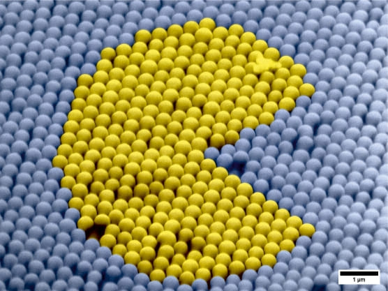
The Rose of Ceres – The image depicts cerium(IV) oxide (CeO₂), which resembles a rose in shape. The title of the image refers to the Roman goddess Ceres – the goddess of agriculture and fertility, after whom the element cerium was named. Cerium(IV) oxide (CeO2) is widely used as a catalytic material in the automotive and chemical industries, as well as in fuel cells, gas sensors and glass polishing. The SEM image was recorded using an FE-SEM FEI Nova NanoSEM 230 and colorized with Adobe Photoshop software. Tungsten Corals – The SEM image reveals tungsten(VI) oxide (WO₃), whose intricate structure resembles the corals of the Great Barrier Reef stretching along the northeastern coast of Australia. Beyond its striking appearance, WO₃ is widely used in gas sensors, smart windows, and advanced electronics because its optical and electrical properties can change in response to light and applied voltage. The SEM image was recorded using an FE-SEM FEI Nova NanoSEM 230 and colorized with Adobe Photoshop software. Photocatalytic PAC-MAN – The SEM image shows a PAC-MAN figure on the surface of a silicon photonic crystal. These materials are nanostructures that enable precise control of light propagation (the light inside this type of structure “moves” much like PAC-MAN in the game) and are widely used in optical fibers, optical sensors, lasers, and advanced telecommunications technologies. The SEM image was recorded using an FE-SEM FEI Nova NanoSEM 230 and colorized with Adobe Photoshop software.
Emilia Wakulska – Wrocław University of Science and Technology
I am currently a second-year doctoral student in the Doctoral School at Wrocław University of Science and Technology, pursuing a degree in materials engineering. My research focuses on the development of luminescent nanoparticles encapsulated in stimulus-responsive polymer coatings.
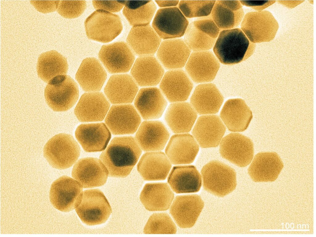
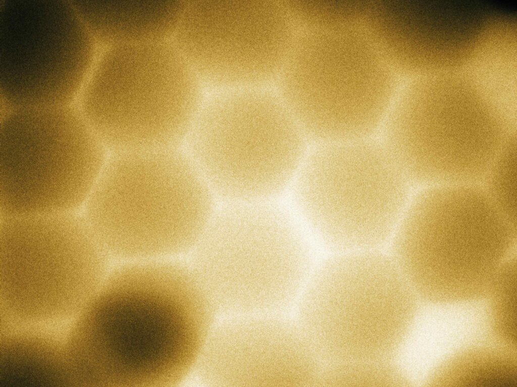
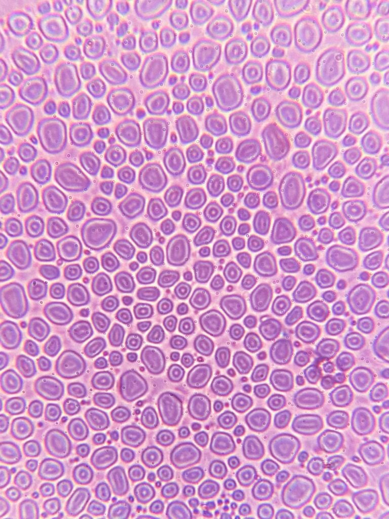
Honeycomb I – Transmission electron microscope (Talos F200i); photo shows NaErF4 nanoparticles photo Honeycomb II – Transmission electron microscope (Talos F200i); photo shows NaErF4 nanoparticles photo Trypophobia nightmare – Optical microscope (Optronic MBL2000); photo shows supramolecular structures – polymersomes
Emilia Wysocka – Łukasiewicz Poznań Institute of Technology
I am a biologist with an artistic soul. On a daily basis, I work as a Biotechnology Specialist at Łukasiewicz Poznań Institute of Technology. In my free time, I enjoy discovering the hidden beauty of the microworld.
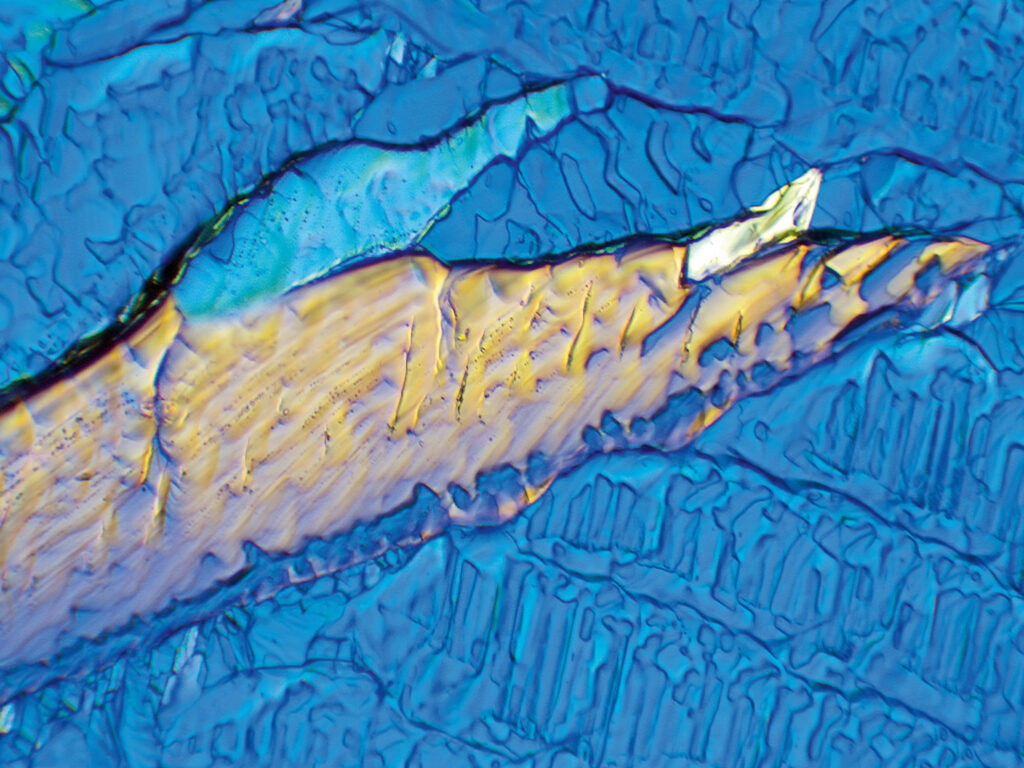
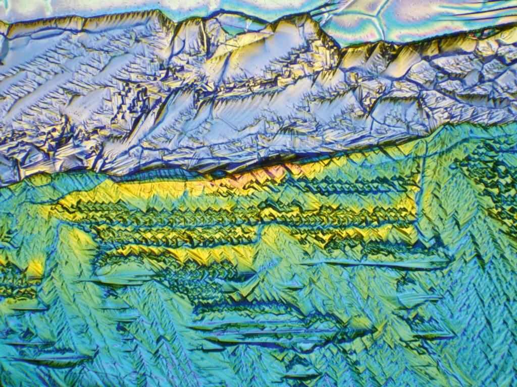
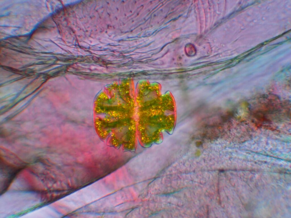
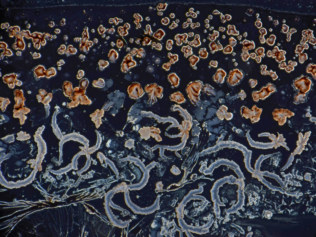
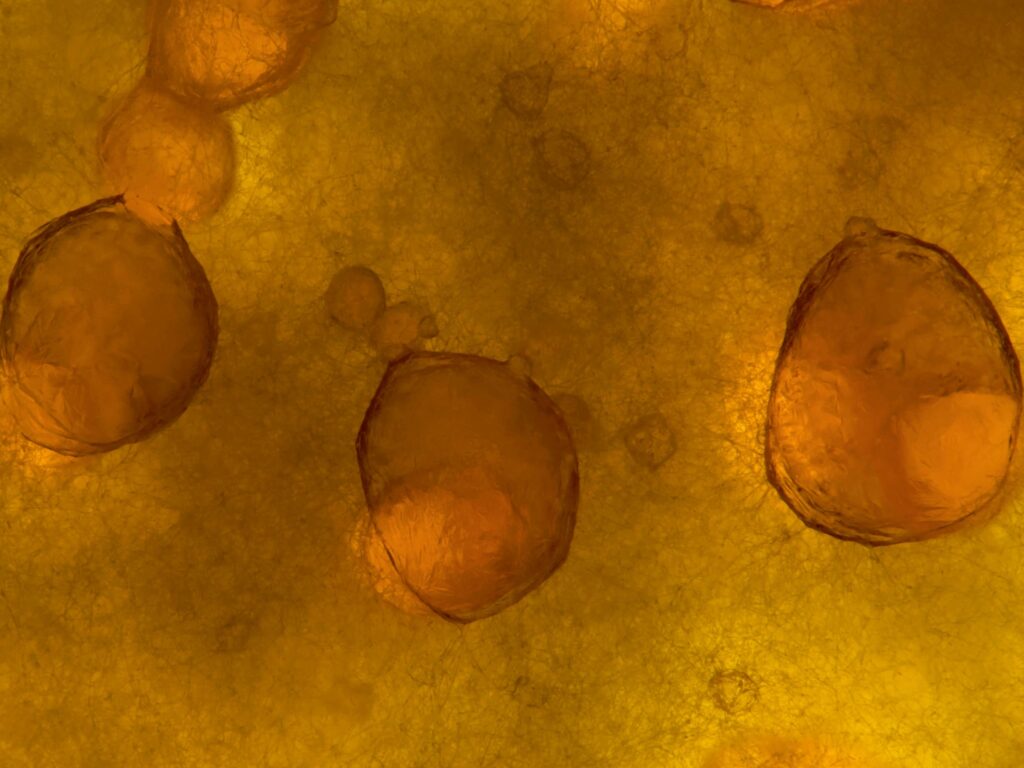
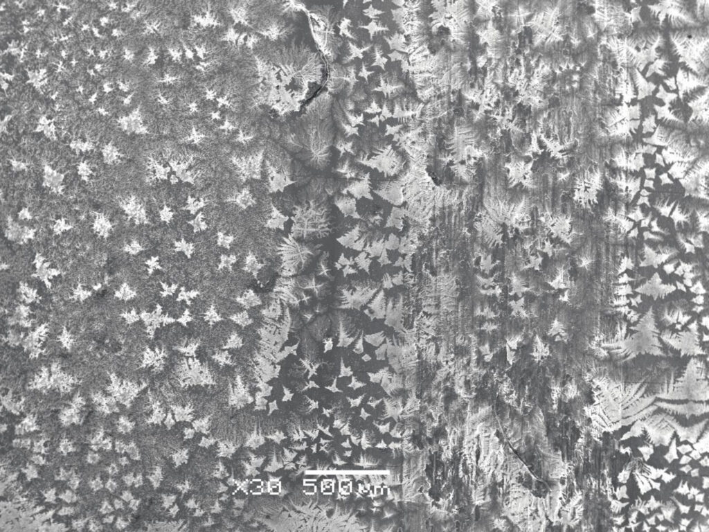
Golden Hand – Taken with the Delta Optical Genetic Pro Trino microscope using polarized light technique. It shows recrystallized aminoacid – beta-alanine with crystal patterns resembling “a golden hand”.Golden Hour – Taken with the Delta Optical Genetic Pro Trino microscope using polarized light technique. It shows recrystallized magnesium sulfate forming patterns of a “golden sunset in the mountains”. Green Gold – Taken with the Delta Optical Genetic Pro Trino microscope using polarized light technique. It shows a shimmering freshwater green alga – Micrasterias truncata. Silver of the Earth – taken with the Delta Optical Genetic Pro Trino microscope using dark-field and polarized light techniques. It shows a recrystallized nitrite-testing reagent for aquariums, which shimmering crystals form a soil-like layer with bluish root-like structures and brownish rock-like forms, resembling an underground biological network. Gold of the Sea – Taken with the Delta Optical Genetic Pro Trino microscope using bright-field technique. It shows brownish spores of an endophytic fungus, isolated from the Wollemi Pine, which resemble sea gold – amber in both shape and color.
Gizella Csire – Institute of Chemistry and Physics, Eszterházy Károly Catholic University
I have a PhD in chemistry – the field of coordination chemistry – Hungary (2017) I currently work in the Department of Chemistry at Eszterházy Károly Catholic University. I am an amateur artist.
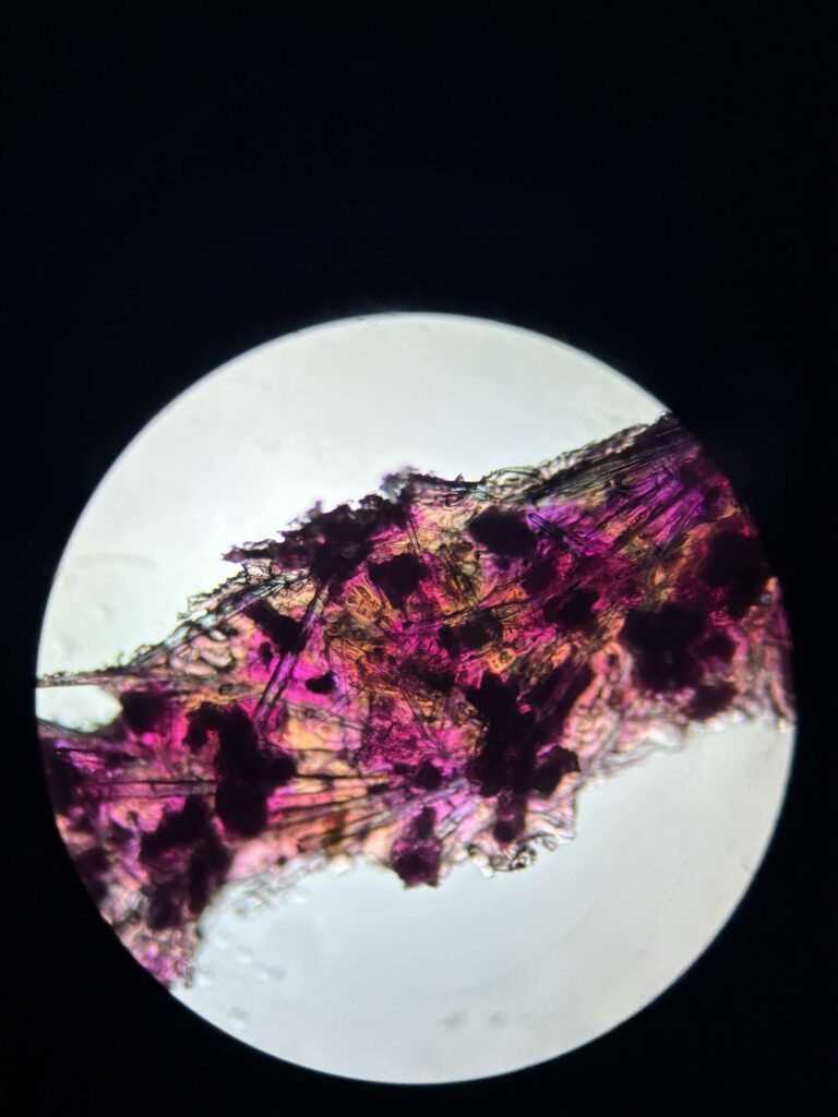
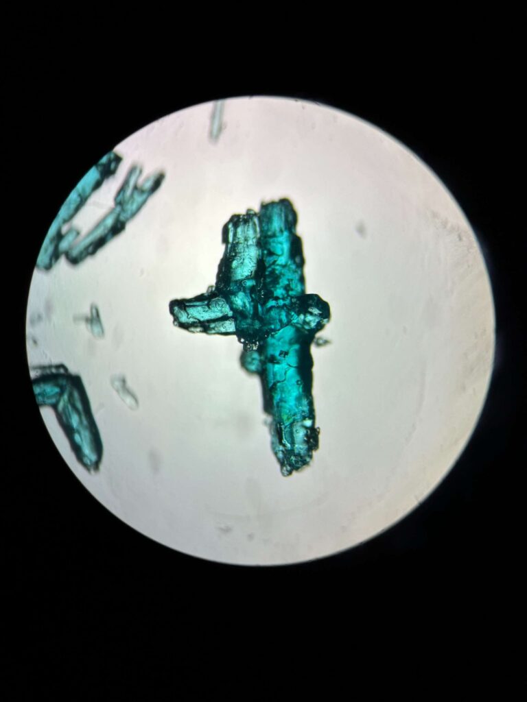
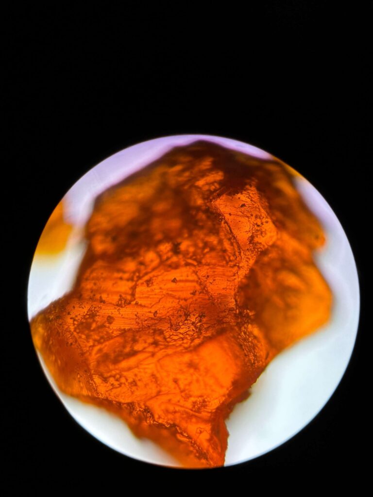
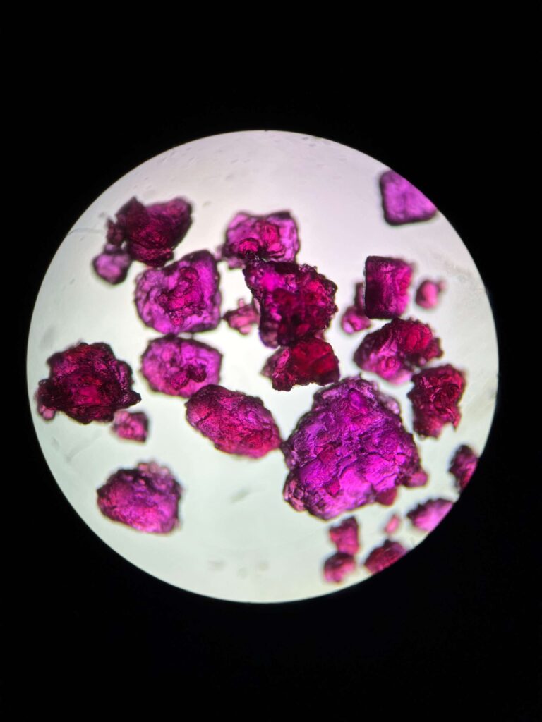
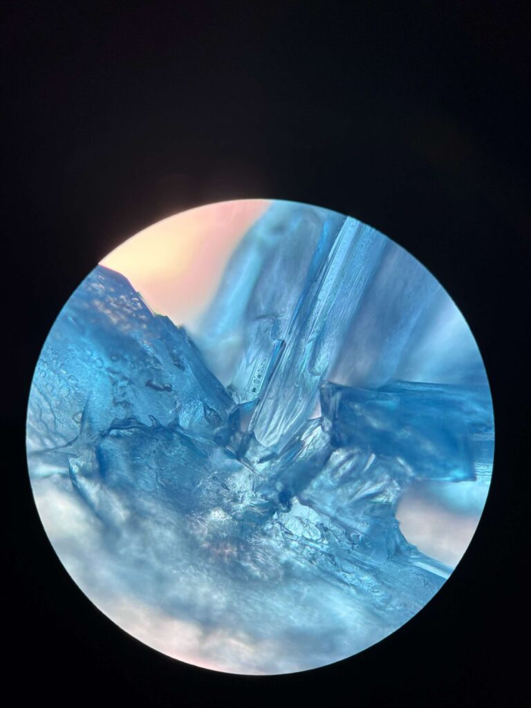
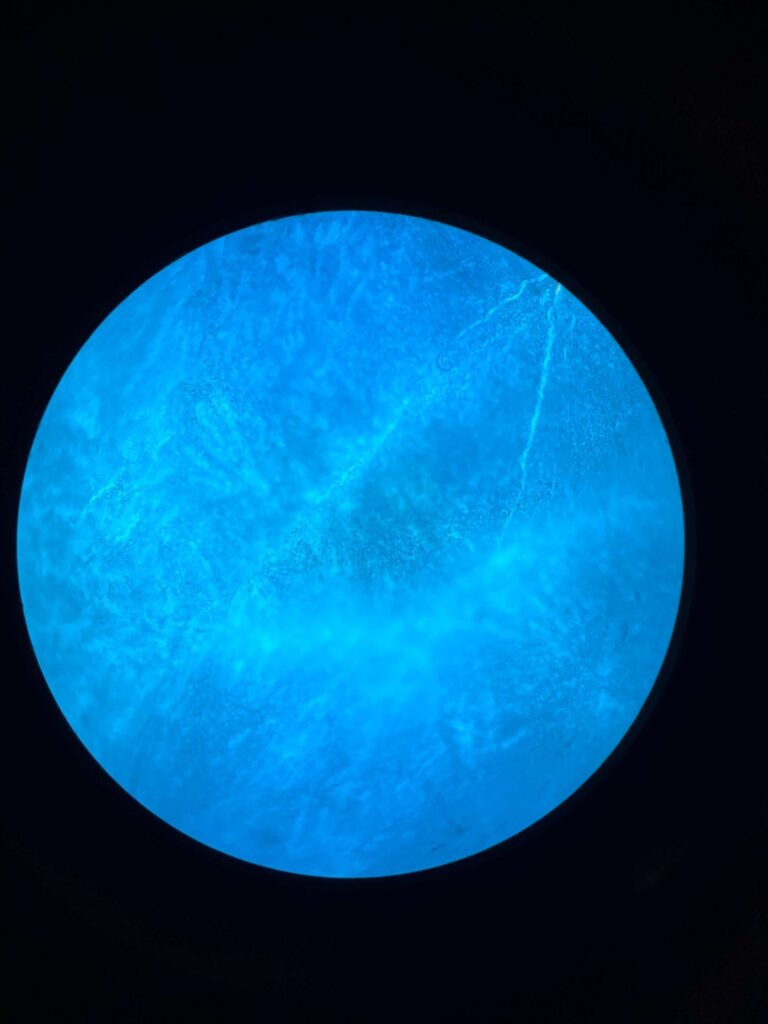
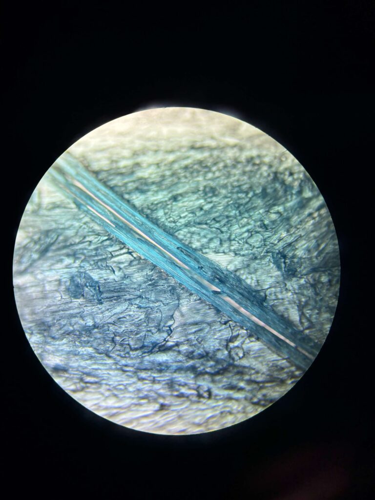
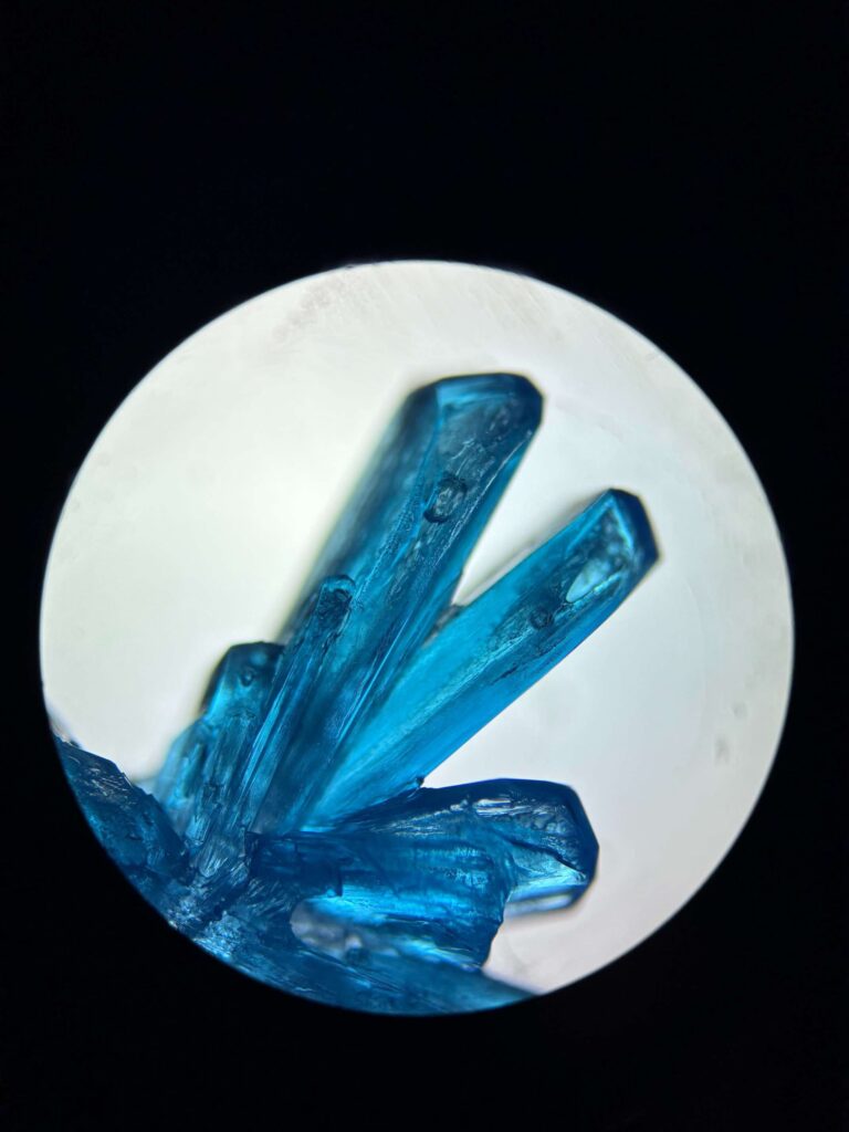
Enchanted by the pink and yellow – FeCl3 – CoCl2, iron(III) chloride – cobalt(II) chloride crystals. Made with Motic/BA210LED Trinocular Microscope (17 mm, 4x/0.10 Zoom) The Blue Duck – CuCl2, copper(II) chloride crystals. Made with Motic/BA210LED Trinocular Microscope (17 mm, 4x/0.10 Zoom) Green Dream NiSO4, nickel(II) sulfate crystals. Made with Motic/BA210LED Trinocular Microscope (17 mm, 4x/0.10 Zoom) Amber – K2Cr2O7, potassium dichromate crystals. Made with Motic/BA210LED Trinocular Microscope (17 mm, 4x/0.10 Zoom) Teddy Bear in the Raspberry Bush – CoCl2 – cobalt(II) chloride crystals. Made with Motic/BA210LED Trinocular Microscope (17 mm, 4x/0.10 Zoom)
Izabella Laszko – Institute of Materials Engineering, Faculty of Science and Technology, University of Silesia in Katowice
I am a PhD student at the International Environmental Doctoral School at the University of Silesia in Katowice. I mainly focus on developing and investigating new biomaterials for potential medical applications, with a particular interest in MMC composites and heterogeneous materials. Beyond science, I enjoy mountain trips, cooking, and learning to play the bass guitar in my free time. I am naturally curious and like bringing creativity into both my research and everyday life.
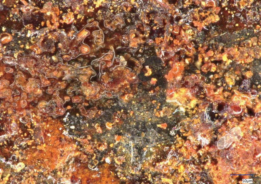
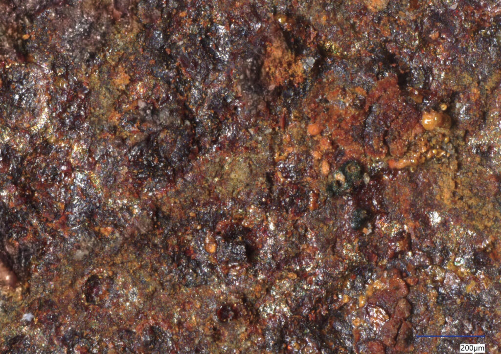
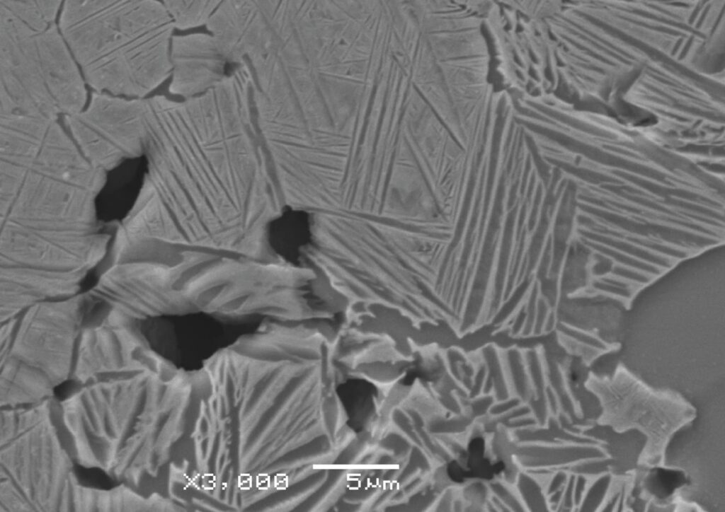
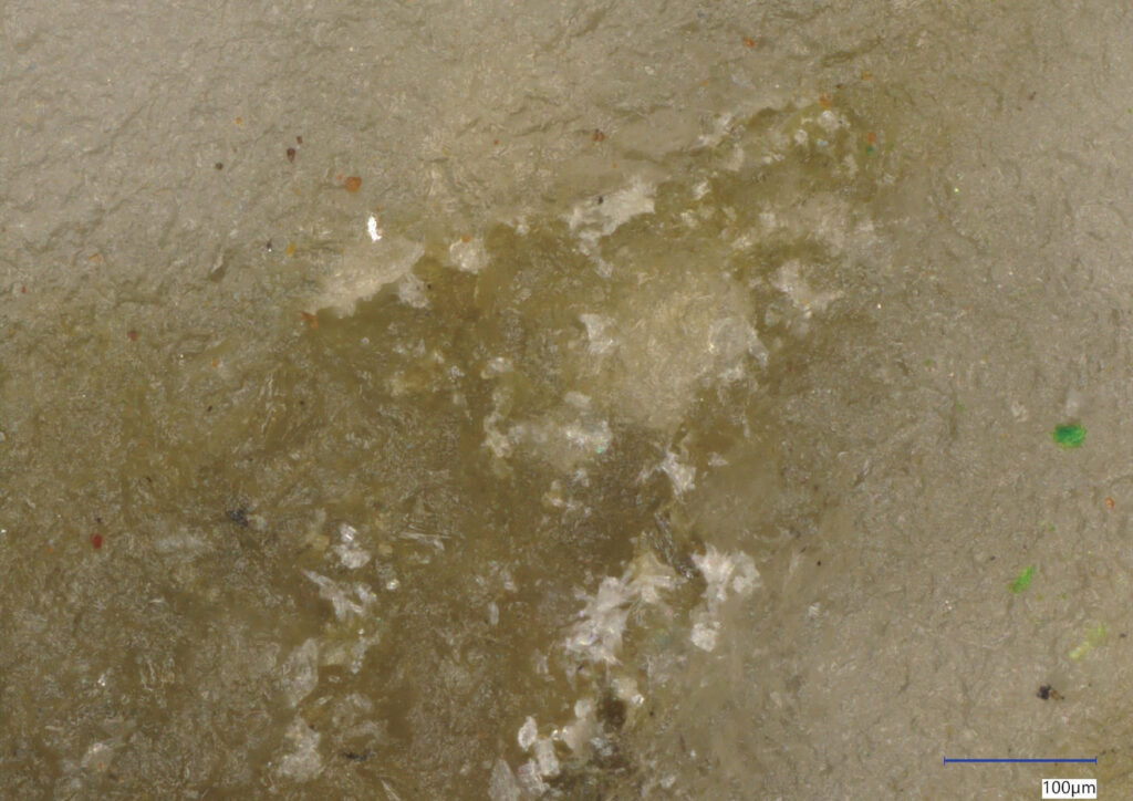
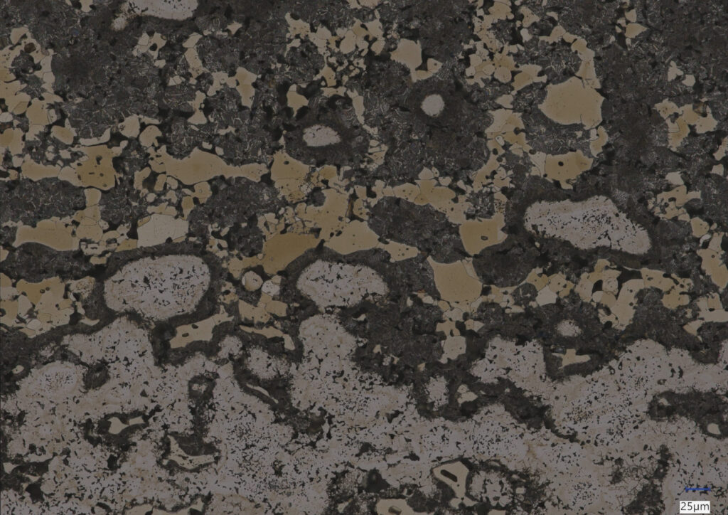
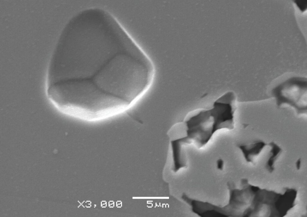
Ordinary and extraordinary corrosion – The image, obtained using a Keyence VHX-X1 digital microscope, presents the surface of a corroded steel wire in microscopic scale. What appears to be ordinary corrosion on a macroscopic scale resembles an abstract work of art under the microscope.What time leaves behind – The image, obtained using a Keyence VHX-X1 digital microscope, presents a close-up view of the surface of a corroded steel washer. Magnification reveals a fascinating complexity of details invisible to the human eye. Cat’s eye – The image, obtained using an Olympus GX1 optical metallographic microscope, presents the microstructure of a titanium matrix composite. The characteristic crack within the reinforcing particle creates the illusion of a cat’s eye hidden in the material. A pigeon enchanted in the microstructure – The image, obtained using a JEOL JSM-6480 scanning electron microscope, presents the microstructure of a titanium matrix composite reinforced with Ti6Al4V alloy particles. The contrast between different areas of the microstructure looks like the profile of a pigeon. Hidden within its own safe zone – The image, obtained using a JEOL JSM-6480 scanning electron microscope, presents the microstructure of a titanium matrix composite reinforced with Ti6Al4V alloy particles. Surrounded by a well-defined boundary, the central structure resembles a form seeking shelter.
Jakub Szarmach – Institute of Fluid-Flow Machinery, Polish Academy of Sciences and Faculty of Applied Physics and Mathematics, Gdańsk University of Technology
I have recently started my journey towards obtaining Master’s degree in Materials Engineering. I have been interested in traditional art forms for quite some time, but with this picture I would like to bridge two things that I love: nanotechnology and art.
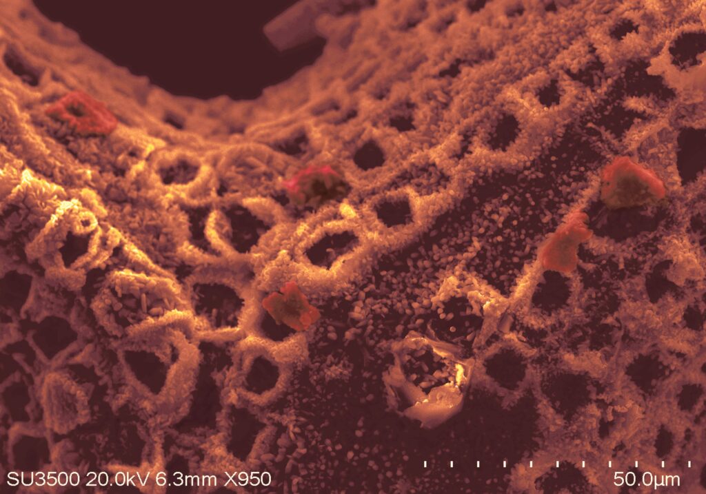
Raspberries from the Sun – Colorized SEM image of anodized thin layers of titanium/titanium oxide sputtered onto both sides of Ficus religiosa leaf skeleton. Picture taken with Hitachi SU3500 SEM.
Jastin Popławski – Wrocław University of Science and Technology
Jastin is PhD candidate in material science at Wrocław University of Science and Technology, his main research area is synthesis and preparation of photonic materials.
Neon Hadal – Fluorescent optical microscopy with Nikon Eclipse Ti2 inverted microscope. Presented micro/nanocomposite – thin film of hollow silica spheres dispersed in polymeric matrix, doped with semiconductor quantum dots.
Joanna Domańska – Jan Długosz University in Częstochowa
I am an MSc student in biotechnology with a strong interest in microbiology, toxicology, and forensic science. As an active member of the MicroLab Microbiology Club, I actively participate in microbiological research and laboratory work. In my free time, I enjoy listening to true crime podcasts, especially Kryminatorium.
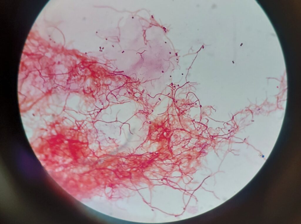
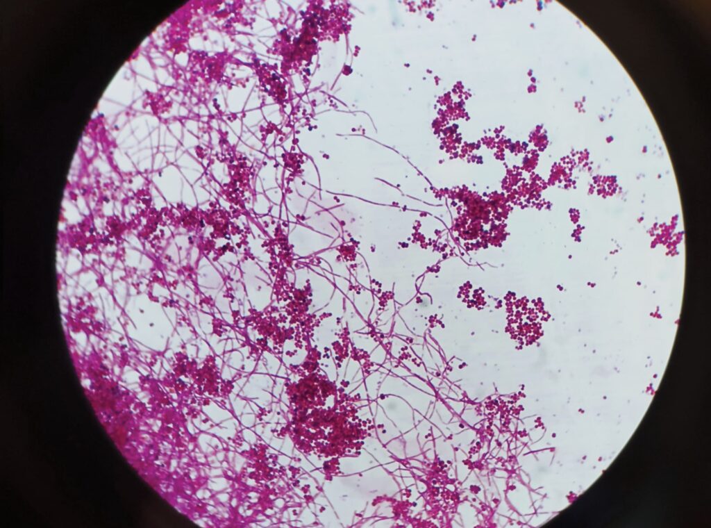
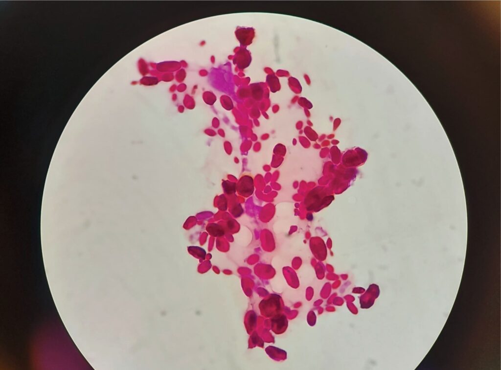
A microscopic forest where every branch tells a story – Captured using an Olympus optical microscope at 1000× magnification with immersion oil. Gram-stained preparation of filamentous Actinomycetes forming delicate branching structures and an intricate microscopic network. The Wi-Fi map inside a Petri dish – Captured using an Olympus optical microscope at 1000× magnification with immersion oil. Gram-stained Actinomycetes creating a dense and dynamic network of branching filamentous structures. Nature really said: add more pink – Captured using an Olympus optical microscope at 1000× magnification with immersion oil. Gram-stained microbial preparation presenting oval-shaped cellular structures arranged in visually striking organic patterns.
Julia Zając – International Environmental Doctoral School, University of Silesia in Katowice
I am a PhD candidate at the International Environmental Doctoral School at the University of Silesia in Katowice, specialising in materials engineering. My research focuses on developing novel zirconium-based metallic biomaterials. I enjoy actively contributing to the academic community and working interdisciplinarily without limiting myself to the technical sciences.
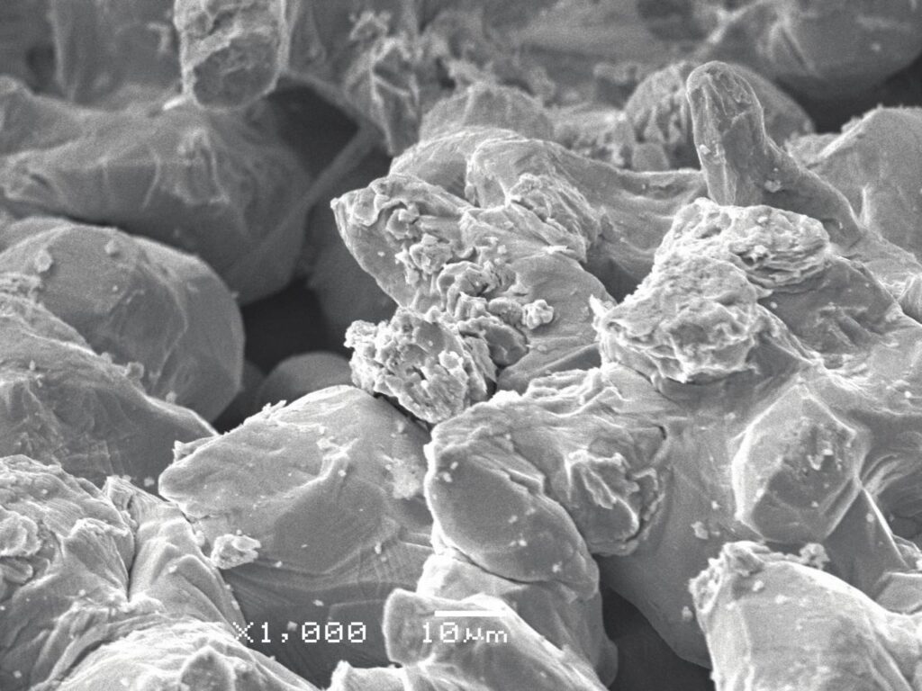
Microworld of proliferation – SEM-SEI image of pure zirconium powder revealing an unexpected structure. The particles exhibit surface features suggestive of localised proliferation, giving the impression of emerging formations. The overall morphology evokes a subtle sense of unease, as if the material itself were undergoing a hidden form of growth within the microworld. Tribological forest – SEM-SEI image of a wear track after tribological testing in Ringer’s solution. The worn surface is covered with localised crystallisation products, creating an unexpected, almost surreal micro-landscape of pine forest. Metal-born Paramecium – A micrograph of an alloy’s microstructure acquired using digital microscope. The grains strikingly resemble a part of a microbiological organism – Paramecium. If you take a closer look you can even notice a contractile vacuole. Cosmos beneath the golden veil – Digital microscope image of cured carbon-based resin revealing a striking, gold-like surface texture. The visual effect arises from differences in light scattering caused by the surface microstructure and variations in reflectivity. As a result, the material forms intricate, flowing patterns reminiscent of cosmic structures, as if a fragment of the universe had been encapsulated within a metallic, golden veil. House No. 20 – Digital microscope image showing an alloy’s microstructure where grains were arranged to form the number “20”. This work is dedicated to my grandparents, both of whom live in places associated with the number 20. Spaces filled with warmth, familiarity, and a strong sense of home.
Konrad Król – Wrocław University of Science and Technology
PhD student at WUST, member of Nanometrology Department
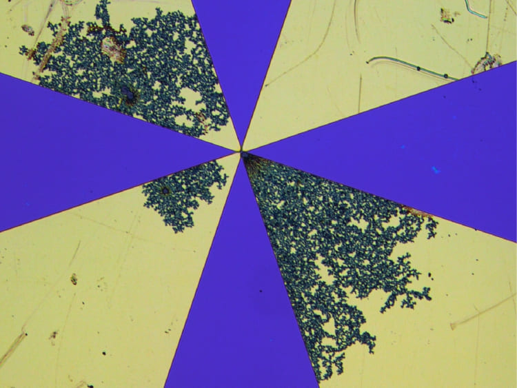
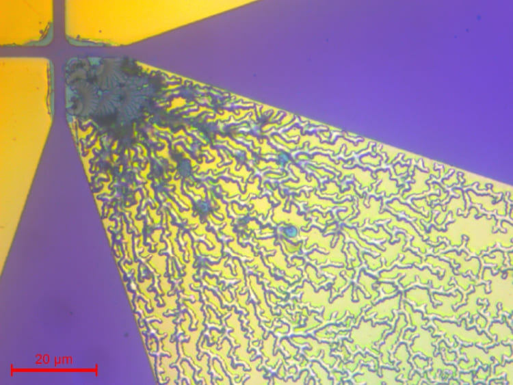
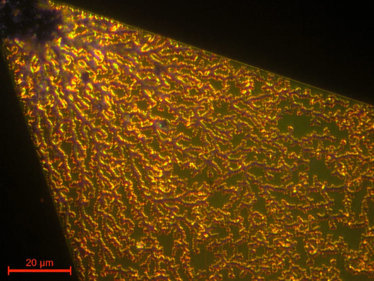
The lace epidemic of gold – The photo shows gold contacts damaged as a result of an electrical breakdown. The image was captured with a Leica optical microscope. The electrical breakdown network – The branched electrical breakdown network is visible across the damaged gold contacts. The image was captured with a Leica optical microscope. Invasion of Golden Lace – The branched electrical breakdown network is visible across the damaged gold contacts. The image was captured with a Leica optical microscope and optical filters.
Krzysztof Wojno – Institute of Fluid-Flow Machinery, Polish Academy of Sciences and Faculty of Applied Physics and Mathematics, Gdańsk University of Technology
Hi, I’m a Materials Engineering master’s student at Gdańsk University of Technology, and I conduct a large part of my research at the Institute of Fluid-Flow Machinery of the Polish Academy of Sciences. I enjoy turning SEM images of my samples into art, as it allows me to share the hidden beauty of the nano-sized world with others. Universe is full of amazing things; look for them, be curious!
Celestial Valley – This colorized image presents the top surface of aligned titania nanotubes covered with thin nickel layer of 5 nm and then treated with the laser beam of 355 nm wavelength with the fluence of 100 mJ/cm2. Titania nanotubes were formed via anodisation of Ti film sputtered onto ITO/glass substrate. The image was taken with scanning electron microscope Hitachi SU3500. Heart On Ice – This colorized image presents the top surface of aligned titania nanotubes covered with thin nickel layer of 5 nm and then treated with the laser beam of 355 nm wavelength with the fluence of 100 mJ/cm2. Titania nanotubes were formed via anodisation of Ti film sputtered onto ITO/glass substrate. The image was taken with scanning electron microscope Hitachi SU3500. Nano Continents – This colorized image presents the top surface of aligned titania nanotubes covered with thin nickel layer of 5 nm and then treated with the laser beam of 355 nm wavelength with the fluence of 100 mJ/cm2. Titania nanotubes were formed via anodisation of Ti film sputtered onto ITO/glass substrate. The image was taken with scanning electron microscope Hitachi SU3500. Oxytocin Breath – This colorized image presents the top surface of aligned titania nanotubes covered with thin nickel layer of 5 nm and then treated with the laser beam of 355 nm wavelength with the fluence of 100 mJ/cm2. Titania nanotubes were formed via anodisation of Ti film sputtered onto ITO/glass substrate. The image was taken with scanning electron microscope Hitachi SU3500.Who’s a Good Boy – This colorized image presents the top surface of aligned titania nanotubes covered with thin nickel layer of 5 nm and then treated with the laser beam of 355 nm wavelength with the fluence of 100 mJ/cm2. Titania nanotubes were formed via anodisation of Ti film sputtered onto ITO/glass substrate. The image was taken with scanning electron microscope Hitachi SU3500.
Maja Janasz – I Liceum Ogólnokształcące im. Stanisława Staszica w Lublinie
I don’t have any achievements, I just really enjoy biology.
Pollen – Pollen of Tulipa under an optical microscope.
Malvína Reiffers Čierniková – Comenius University in Bratislava, Faculty of Natural Sciences, Department of Soil Science
I am a researcher at the Department of Soil Science, Faculty of Natural Sciences, Comenius University in Bratislava, where I have been professionally active since my undergraduate studies. My primary expertise lies in wetland ecology and palynology, with a specialized focus on pollen determination and paleopalynological vegetation reconstruction. While my current research investigates the intersection of ecological factors and plant growth through functional traits, palynology remains my true passion, leading to my long-term vision of creating a comprehensive photo atlas of pollen grains based on my original microphotography.
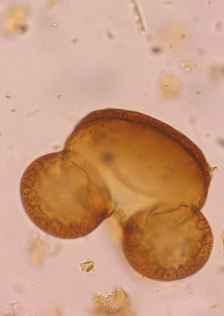
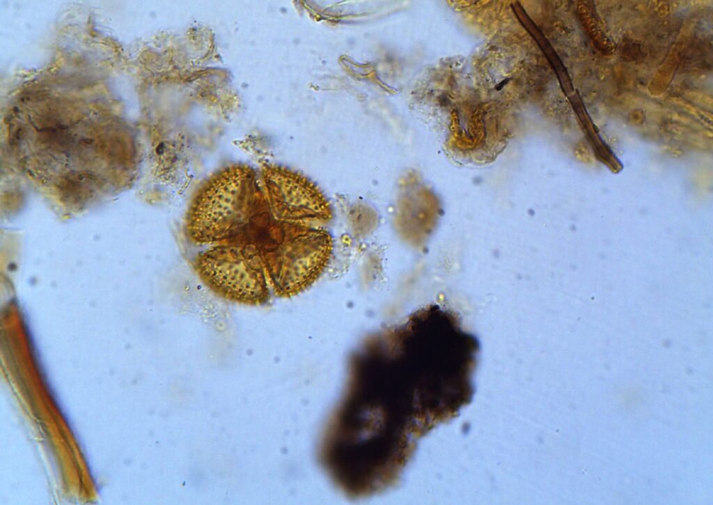
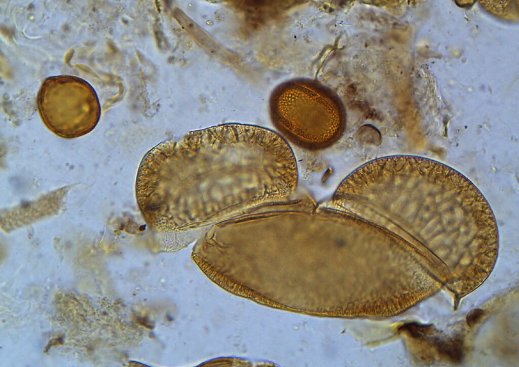
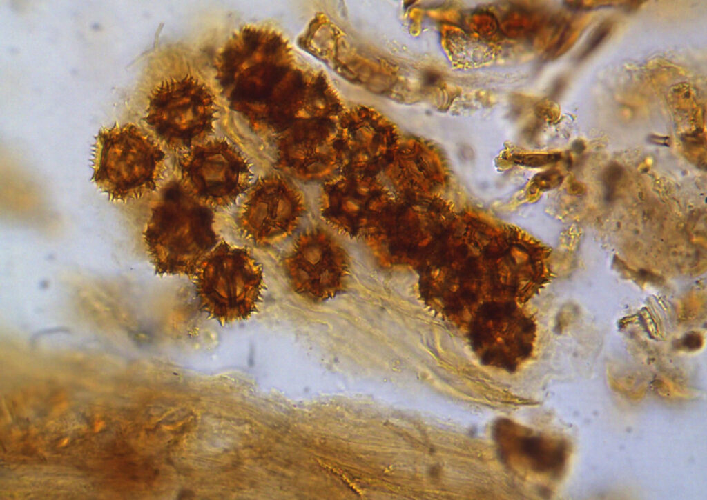
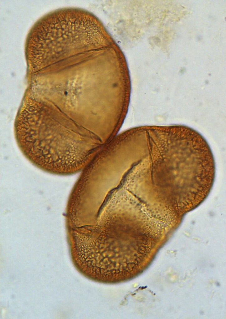
The Micro-Mickey – Captured with an OPTIKA B-510BF trinocular light microscope, this image shows a single bisaccate pollen grain of Abies alba. The distinct air bladders, which aid in wind dispersal, create a silhouette reminiscent of iconic pop-culture figures. Unidentified Flying Object – This image, taken using an OPTIKA B-510BF trinocular light microscope, features a pollen grain of Drosera with a striking four-fold symmetry. The intricate surface ornamentation and geometric central structure highlight the natural complexity of microscopic plant reproductive cells. The Micro-Archipelago – A diverse cluster of pollen grains – Fagus sylvatica, Fagopyrum esculentum, Abies alba – and organic debris is shown here, captured with an OPTIKA B-510BF trinocular light microscope. The composition illustrates the variety of shapes and sizes found within a single sediment sample, showcasing a miniature biological landscape. Viral Gossip – Captured through an OPTIKA B-510BF trinocular light microscope, this micrograph reveals a dense colony of echinate (spiky) pollen grains from the Asteraceae family. The sharp exterior spines are evolutionary adaptations designed to adhere to the bodies of pollinating insects. The Big Bang Theory – This detailed view of two bisaccate pollen grains of Picea abies in close contact was achieved using an OPTIKA B-510BF trinocular light microscope. The image focuses on the delicate reticulate patterns of the saccate structures used for aerodynamic stability during flight.
Małgorzata Małecka – Institute of Low Temperature and Structure Research, Polish Academy of Sciences
dr hab. inż. Małgorzata Małecka prof. INTIBS A chemist, a specialist in transmission electron microscopy. Her research focuses on the synthesis and analysis of nanomaterials and hierarchical catalysts based on cerium oxide.
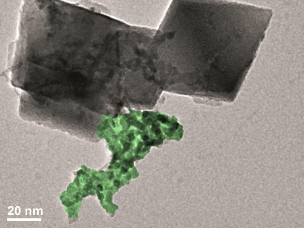
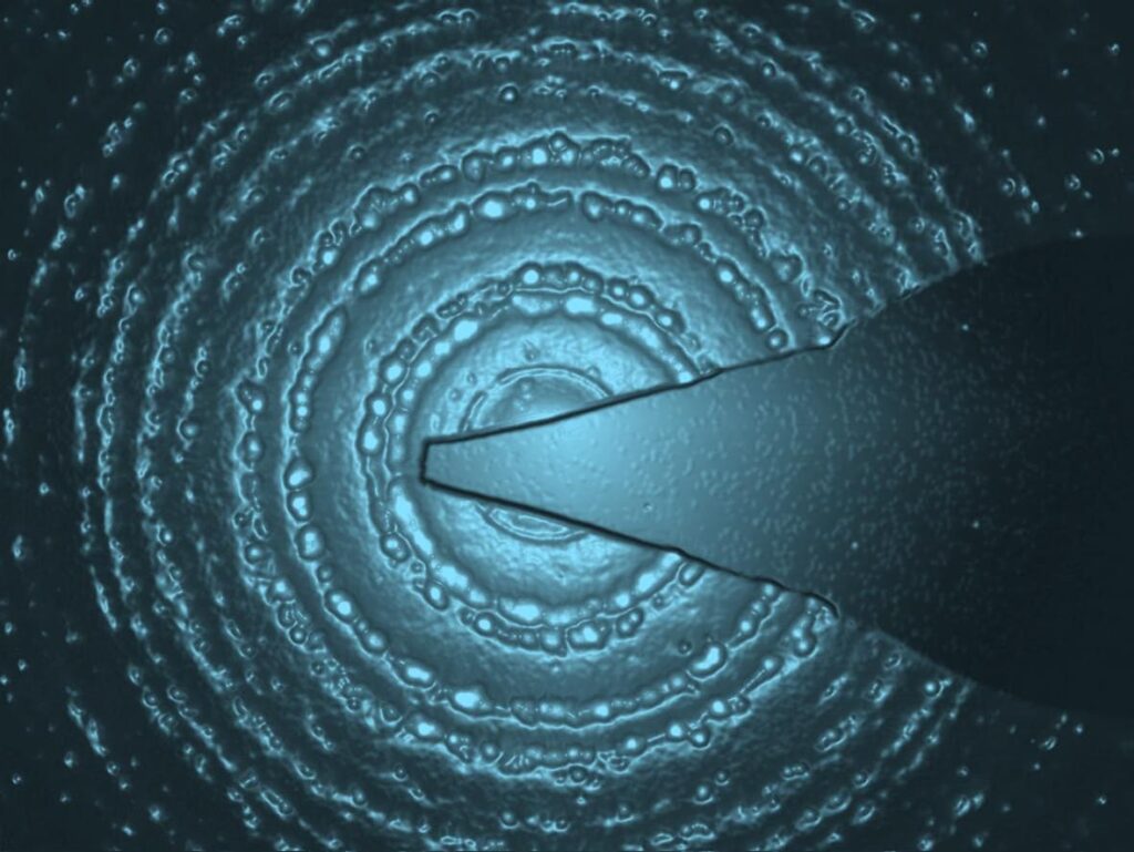
on the shoulders of nano-giants – transmission electron microscope Philips CM20 SuperTwin, ruthenium oxide nanoparticles deposited on octahedral CeO2 crystallites drop of symmetry – transmission electron microscope Philips CM20 SuperTwin, electron diffraction pattern of a Ru/CeO2 sample
Marek Czyszczoń, Dominika Benkowska-Biernacka – Wrocław University of Science and Technology
Marek is a first year PhD student working on the synthesis and characterization of carbon nanodots. He is a part of the Biophotonics Group at Wrocław University of Science and Technology. Dominika Benkowska-Biernacka works on biologically relevant lipidic mesophases doped with fluorescent carbon nanodots as part of her doctoral research at WUST. She completed research internships at École Normale Supérieure Paris-Saclay, KU Leuven, Kent State University, and University of Colorado Boulder dedicated to the synthesis of gold nanoparticles and the application of advanced microscopy techniques for studying lyotropic liquid crystals.
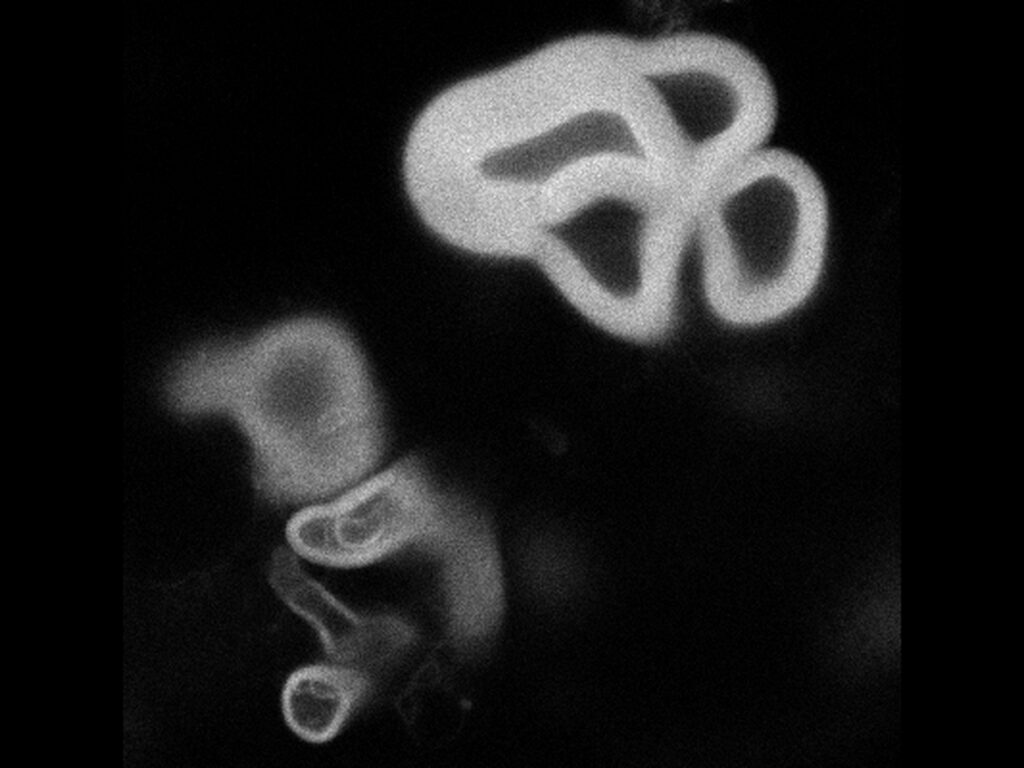
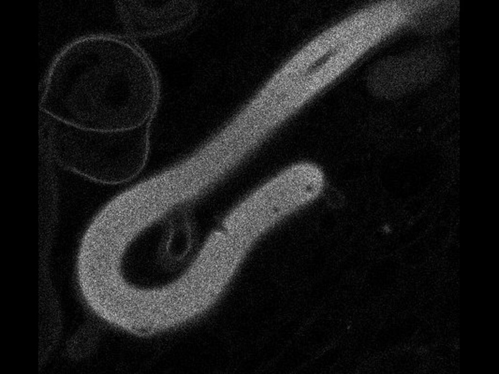
Four-leaf clover set on fire with a lighter by an anonymous hand – The photo depicts phospholipid-based myelin figures doped with carbon nanodots. It was taken using an Aberrior Steadycon STED microscope operating in the confocal mode. A worm going back home because she left a pot on the stove – The photo depicts phospholipid-based myelin figures doped with carbon nanodots. It was taken using an Aberrior Steadycon STED microscope operating in the confocal mode.
Mária Matejová – Comenius University in Bratislava
I am a Master’s student at the Department of Animal Physiology and Ethology at Comenius University in Bratislava, focusing on the effects of prenatal environmental factors on offspring development. Alongside research, I enjoy collecting and exploring new hobbies.
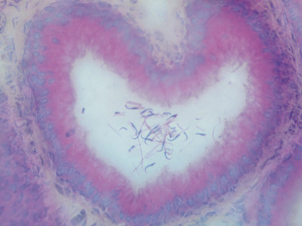
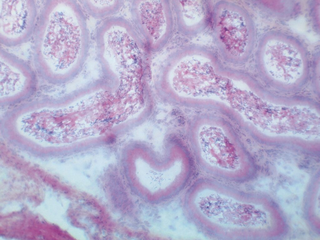
Spheartmia – Rat epididymal duct, 40x magnification, H&E staining, imaged using a Bresser Researcher Trino compound light microscope. Heart in the Right Place – Heart-shaped duct of the rat epididymis, 10x magnification, H&E staining, imaged using a Bresser Researcher Trino compound light microscope.
Martyna Szyszka – Wrocław University of Science and Technology
In my research work, I focus on 3D printing biomaterials containing microalgae. The study was financed by NCN (Grants No. 2023/49/B/ST8/03834).
Blood vessel – The photos show the structure of a 3D-printed hydrogel containing microalgae, shown from different perspectives. The photo was acquired using a Leica DMi8 manual microscope with fluorescence imaging. Algal highway – The photos show the structure of a 3D-printed hydrogel containing microalgae, shown from different perspectives. The photo was acquired using a Leica DMi8 manual microscope with fluorescence imaging. In a microalgae galaxy far, far away… – The photos show the structure of a 3D-printed hydrogel containing microalgae, shown from different perspectives. The photo was acquired using a Leica DMi8 manual microscope with fluorescence imaging. Scar – The photos show the structure of a 3D-printed hydrogel containing microalgae, shown from different perspectives. The photo was acquired using a Leica DMi8 manual microscope with fluorescence imaging. Inner plant sunset – A hydrogel disc containing microalgae. The photo was acquired using a Leica DMi8 manual microscope with fluorescence imaging.
Mikołaj Matys – III Liceum Ogólnokształcące im. Unii Lubelskiej w Lublinie
I’m a student of 3 LO Lublin, passionate about biology and tardigrades.
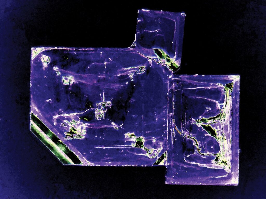
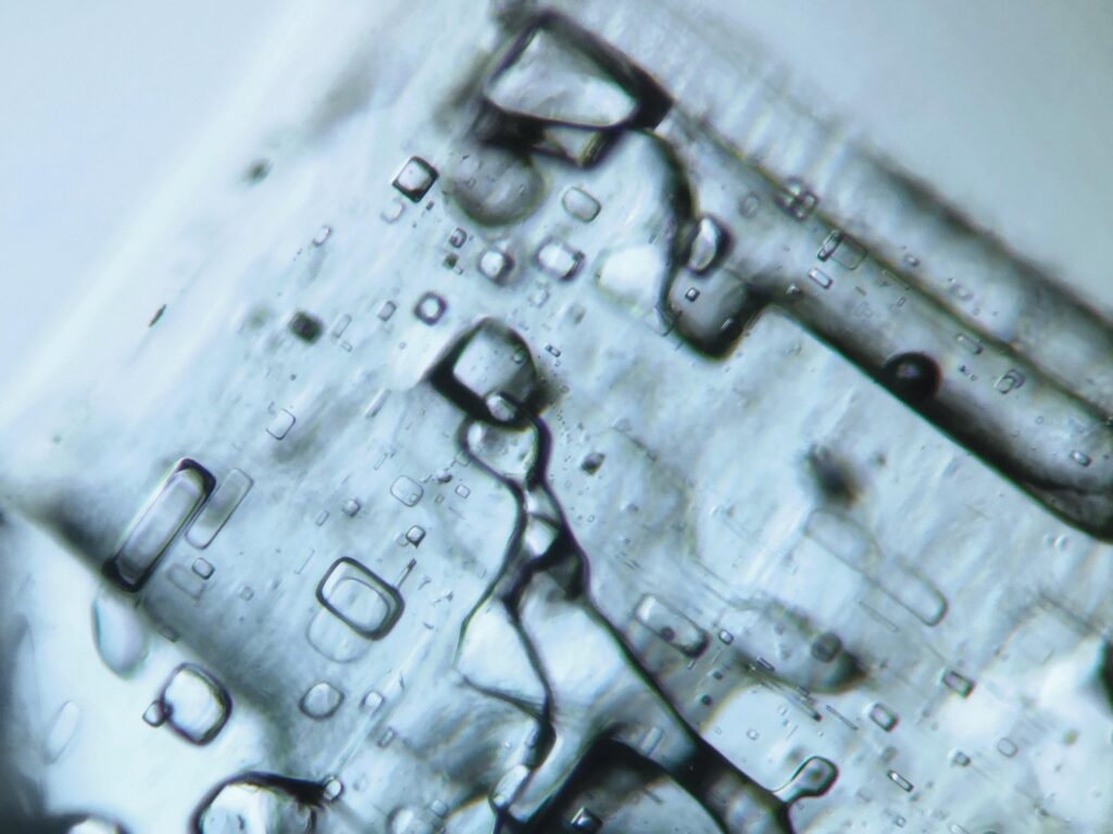
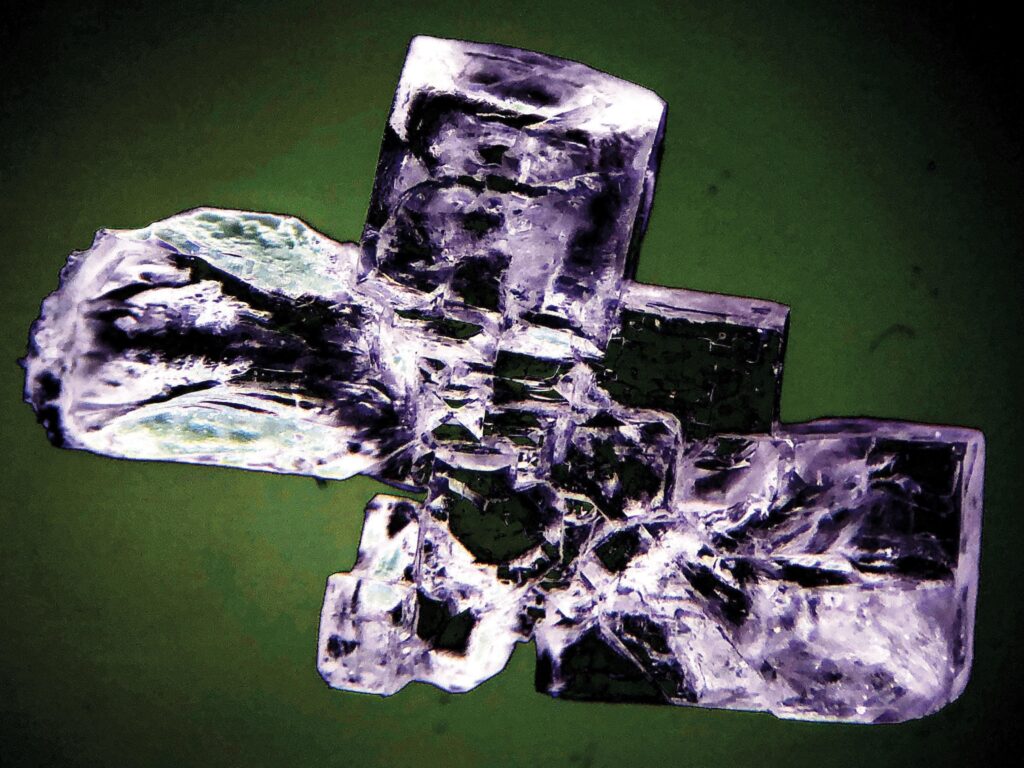
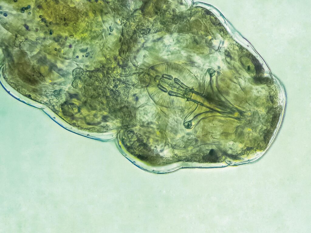
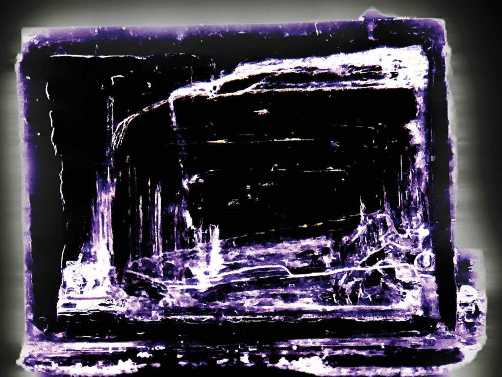
A pinch of salt – stereo microscope, salt crystal (computer-colored) Bubbles escape – stereo microscope, oxygen bubbles trapped in a salt crystal (computer-colored) Glass brick – stereo microscope, salt crystal (computer-colored) Hallo World – optic microscope, 400x magnification, tardigrade – Hypsibius dujardini Infinite loop – stereo microscope, linear structures inside a salt crystal (computer-colored)
Nandhini Ravindran – Physics of Nanostructured Materials Group, Faculty of Physics, University of Vienna
I am a young microscopist. I explore atoms using the super cool Nion UltraSTEM 100 (Scanning Transmission Electron Microscope). As a doctoral student in Physics, I am learning a lot about the world of physics and would like to eventually contribute in a meaningful way towards the building of a sustainable, safe and friendly society/world.
The Alps Climbers Photo – The aberration-corrected ultra-high vacuum scanning transmission electron microscope Nion UltraSTEM 100 was used to capture the image. It is a dark field image of MoS2 on Graphene heterostructure on a transmission electron microscope grid (TEM). In this image, we can see what looks like climbers going up on the Alps. Hey Dude! Don’t try to pluck me! I’m a nano flower – A tabletop low-voltage transmission electron microscope (LVEM5 Delong instruments) was used to capture the image. The freestanding hexagonal boron nitride on TEM grid shows a nanoflower. I’m a peacock of the nano world – Nanopatterns of peacock showed up on the freestanding hBN monolayer on a TEM grid. The image was captured on a tabletop low-voltage transmission electron microscope (LVEM5 Delong instruments). Are you a disco dancer? I am a disco ball! – A tabletop low-voltage transmission electron microscope (LVEM5 Delong instruments) was used to capture a freestanding hBN on TEM grid that shows up as a disco ball. Arctic Aurora blooms – Arctic Aurora bloomed while trying to focus the sample of hBN on a TEM grid at the tabletop low-voltage transmission electron microscope (LVEM5 Delong instruments).
Olga Rebain – Nicolaus Copernicus University in Toruń
I am a student of Medicinal Chemistry. My academic background provides me with a strong foundation in both biological and chemical sciences, enabling me to take an interdisciplinary approach to these fields. I am particularly interested in the design of modern therapeutics and the development of innovative drug delivery systems, with a specific focus on pharmacogenomics.
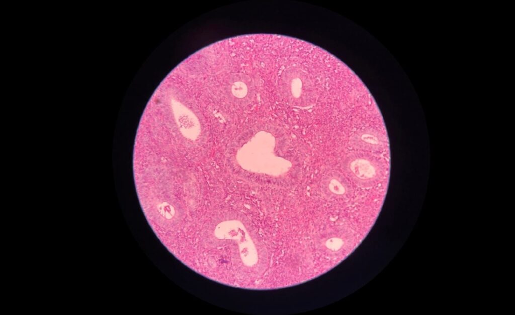
Love Story – This micrographic image presents a histological section of the endometrium in the proliferative phase. The photograph was captured using a light microscope via the afocal technique. Standard hematoxylin and eosin staining was applied. Numerous endometrial glands are visible; the focal point of the image is a gland with an atypical, heart-shaped cross-section -a rare example of natural pareidolia within biological structures.
Orsolya Bogdányi-Fekete – Institute of Chemistry and Physics, Eszterházy Károly Catholic University
I’m a laboratory technician. I help the university educators and students. In my freetime, I adore painting by numbers and hiking with my husband.
Frozen Dream – Recrystallized copper-sulfate. Made with Motic/BA210LED Trinocular Microscope (10X/0.25 zoom, 6.4mm) The Cu-L Planet – Recrystallized copper-sulfate. Made with Motic/BA210LED Trinocular Microscope (10X/0.25 zoom, 6.4mm) The Cuprate Canyon – Recrystallized copper-sulfate. Made with Motic/BA210LED Trinocular Microscope (10X/0.25 zoom, 6.4mm) Peace and Love – Recrystallized copper-sulfate. Made with Motic/BA210LED Trinocular Microscope (17 mm, 4x/0.10 Zoom) Precious Peacock – Recrystallized copper-sulfate. Made with Motic/BA210LED Trinocular Microscope (17 mm, 4x/0.10 Zoom)
Ovidiu-Ioan Agârbicean – Babeș-Bolyai University
I am a passionate medical physics student and an aspiring theoretical physicist. My work spans Ising chain models, radiotherapy and radiation protection, medical imaging techniques, and, last but not least, partial differential equations. For me, microscopy is a beautiful way to bridge science with another deep interest of mine: art.
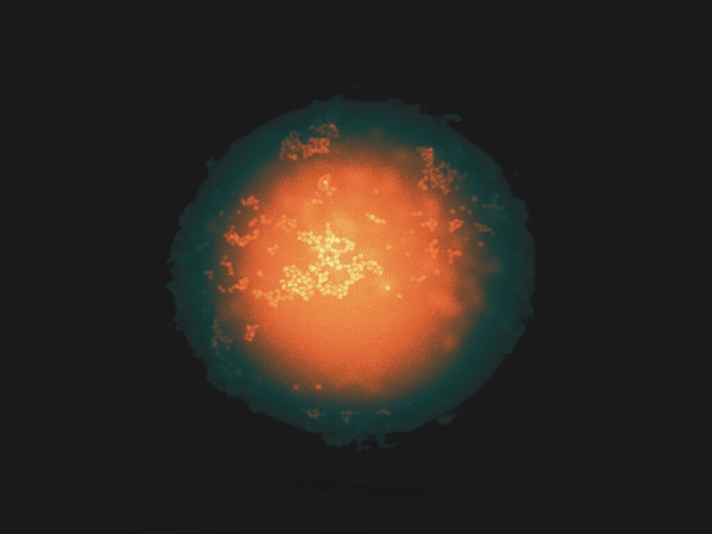
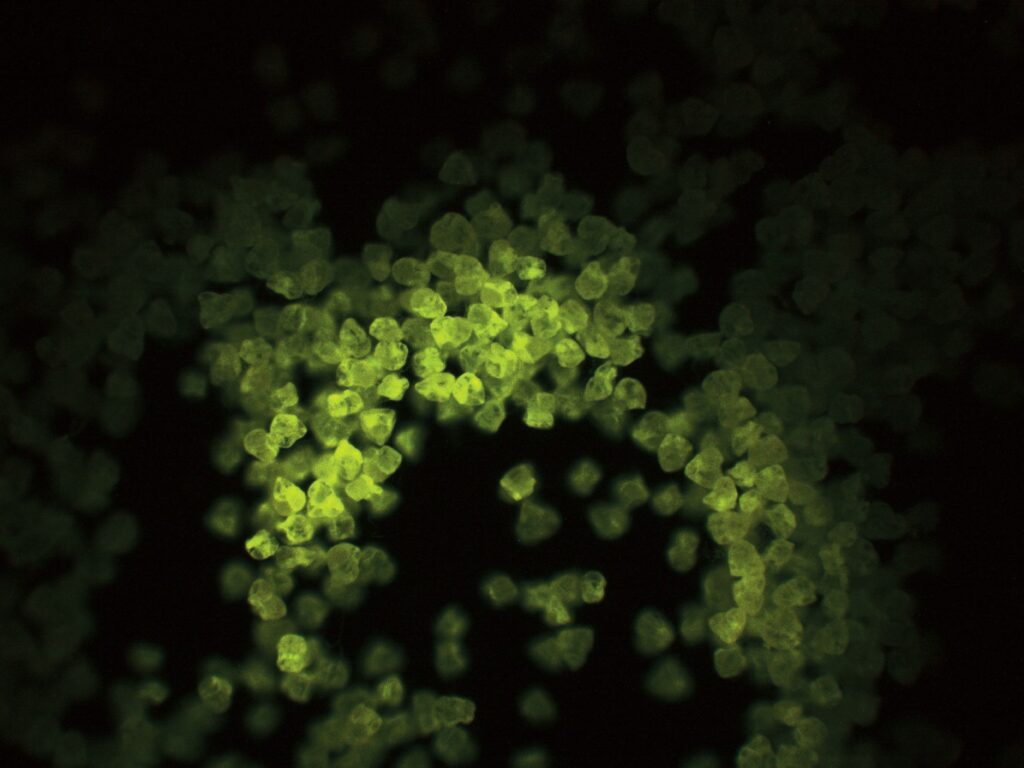
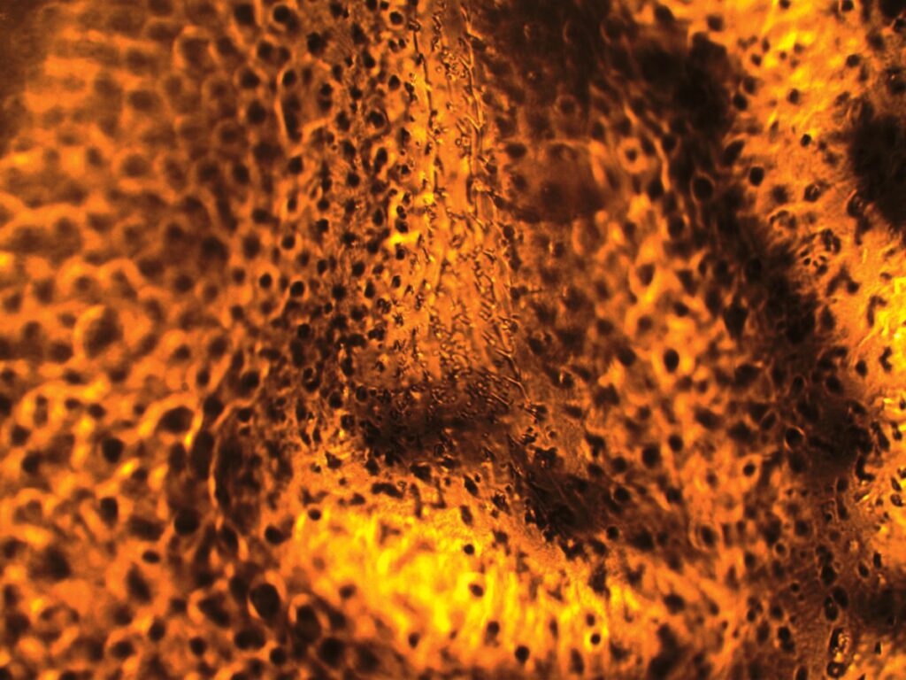
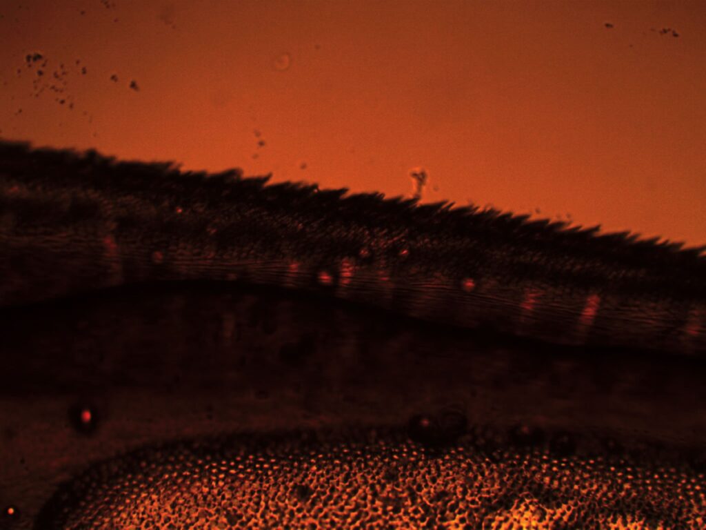
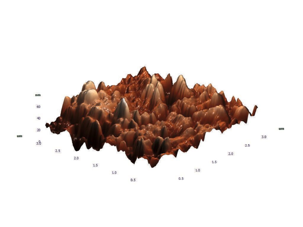
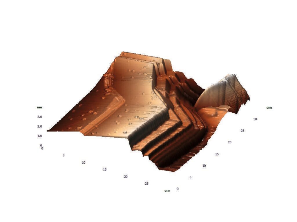
Pollen Nebula – Fluorescence microscopy of pollen grains. (10X, exposure 430ms, 100 gain) A cloud of glowing pollen particles appears suspended in darkness, resembling a microscopic constellation or biological nebula. The image highlights the natural fluorescence and clustered morphology of pollen. Demogorgon Bloom – Optical transmission microscopy of a beetle wing. (40X, exposure 30ms, 20 gain) The porous texture and fiery orange tones create the impression of an open organic mouth, almost like a Demogorgon-like structure (well-known D&D and Stranger Things character). What is biologically a beetle wing becomes, under the microscope, a dramatic landscape of cavities and ridges. Meadow by the Microscopic Lake – Optical transmission microscopy of a beetle wing. (10X, exposure 30ms, 20 gain) This image transforms the edge of a beetle wing into an imagined landscape: a dark meadow, a small reflective lake, and fields reminiscent of wheat under a warm sky. The fine wing structures resemble grass, soil, and distant vegetation. Bubbly Mesa – Atomic force microscopy of recrystallized Terapia paracetamol after dilution in ethyl alcohol. Atomic force microscopy of recrystallized paracetamol after dilution in ethyl alcohol. An AFM landscape filled with rounded peaks and clustered formations, resembling a surreal desert mesa shaped by erosion. The nanoscale topography creates the impression of bubbling terrain rising from the dried paracetamol film. The Grand Atomic Canyon – Atomic force microscopy of recrystallized Zentiva paracetamol after dilution in ethyl alcohol. A smoother AFM landscape with terraces, ridges, and layered crystalline steps spread across the surface. The image evokes a surreal desert plateau reminiscent of the Grand Canyon in Colorado, where sharp nanoscale cliffs and smooth plains emerge from the recrystallized paracetamol film.
Paweł Filipek – II Liceum Ogólnokształcące im. Hetmana Jana Zamoyskiego w Lublinie
I am a student at the II High School in Lublin and I am interested in biology.
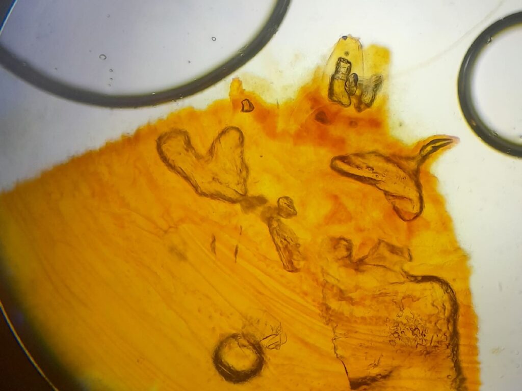
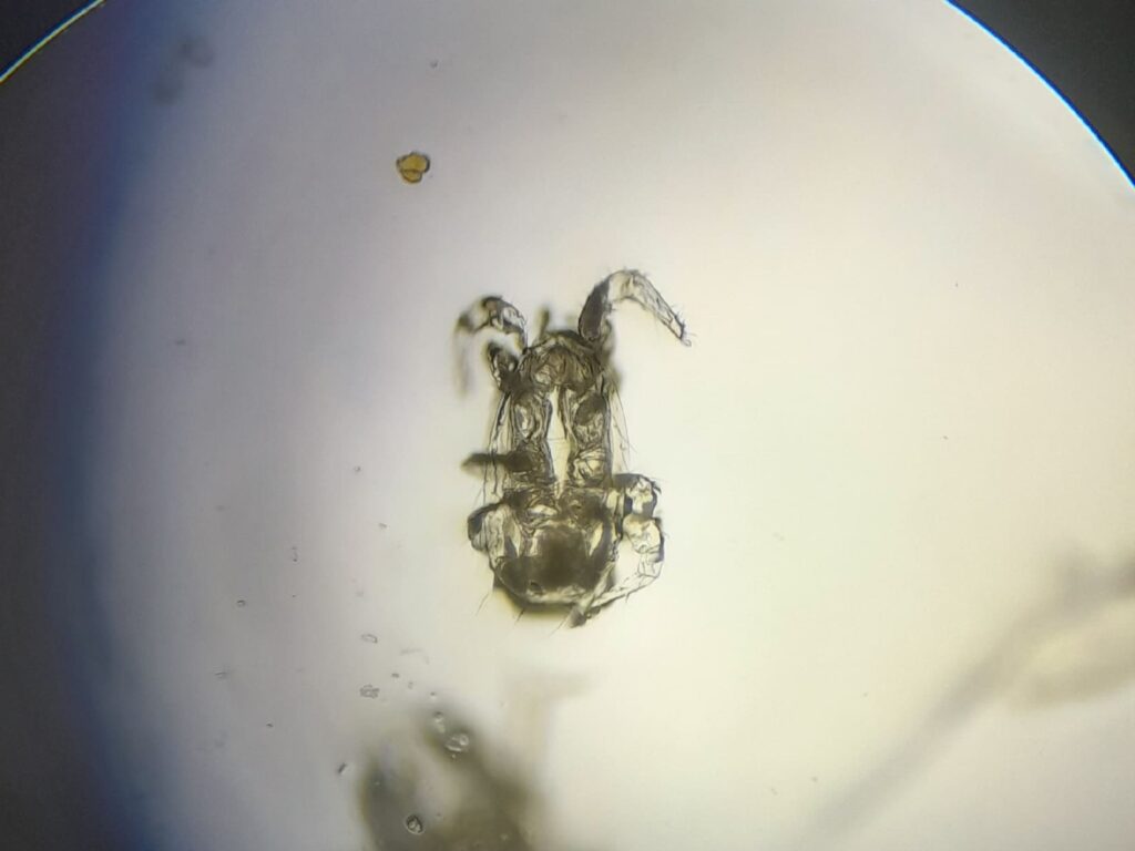
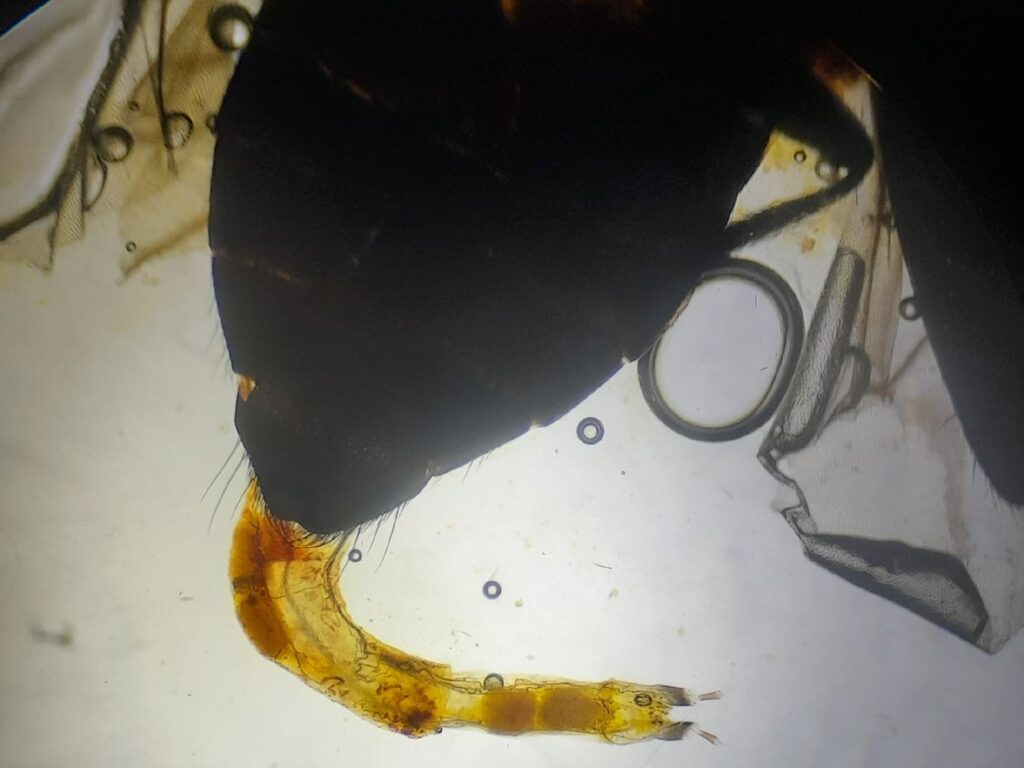
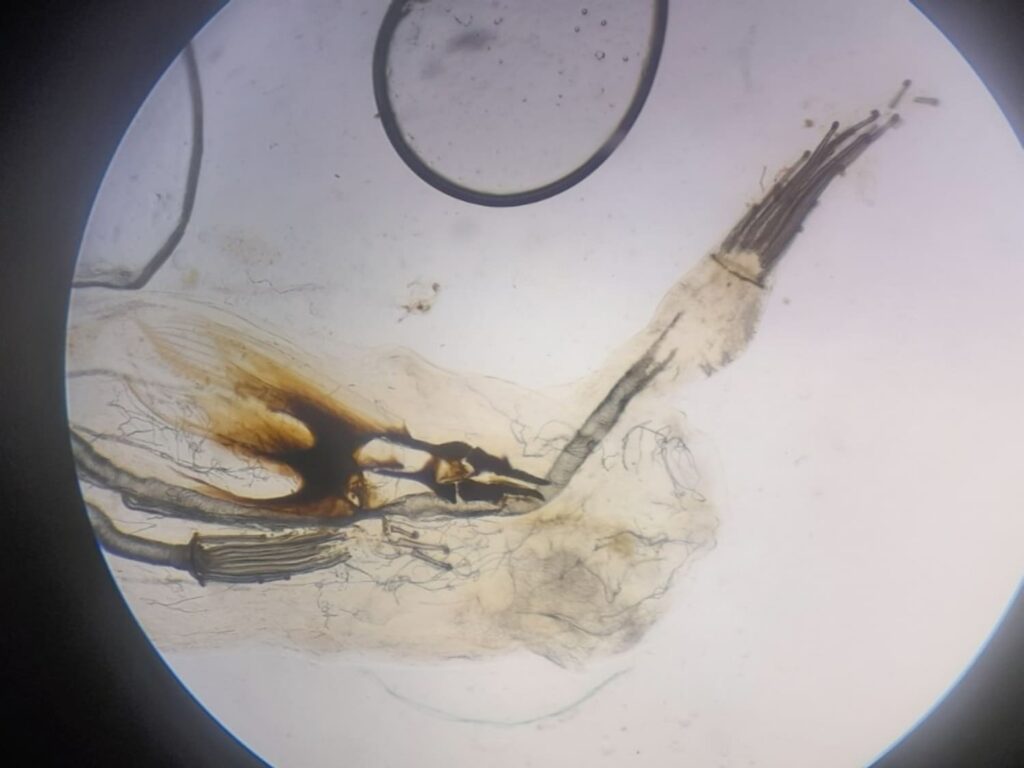
Remnants of the larval mouthparts in the pupa of Drosophila melanogaster – The photo was taken with an optical microscope and shows Remnants of the larval mouthparts in the pupa of Drosophila melanogaster. Preparat Amblyseius swirskii – The photo was taken with an optical microscope and shows Amblyseius swirskii. Copulatory apparatus of the male Brassicogethes aeneus – The photo was taken with an optical microscope and shows copulatory apparatus of the male Brassicogethes aeneus. Malpighian tubules in the larva of Cydia pomonella – The photo was taken with an optical microscope and shows Malpighian tubules in the larva of Cydia pomonella.
Piotr Kuich – Wrocław University of Science and Technology
PhD student in Materials Engineering, affiliated with the Department of Physical and Quantum Chemistry and the Institute of Advanced Materials at the Faculty of Chemistry, Wrocław University of Science and Technology.
The Ocean Depths – The image was acquired using Transmission Electron Microscopy (Thermo Fisher Scientific Talos F200i, 200 kV) in Scanning Transmission Electron Microscopy (STEM) mode with High-Angle Annular Dark Field (HAADF) imaging. It represents ZnGa₂O₄:Cr³⁺ persistent luminescence nanoparticles encapsulated in L-α-phosphatidylcholine-based vesicular type nanocarrier, stabilized with the mild surfactant cocamidopropyl betaine.
Réka Joharchy – Institute of Chemistry and Physics, Eszterházy Károly Catholic University
I’m a laboratory technician. My interests are doing spectacular chemistry experiments and growing crystals of metal salts. In my free time, I like reading fantasy stories and playing with my cats and dogs.
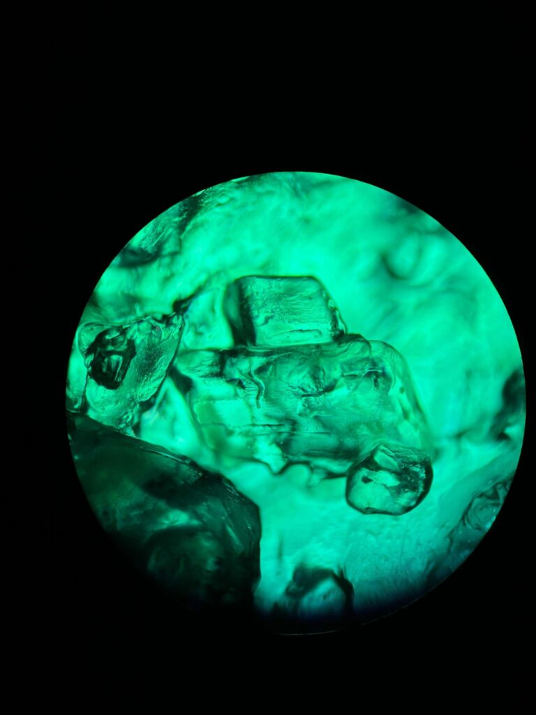
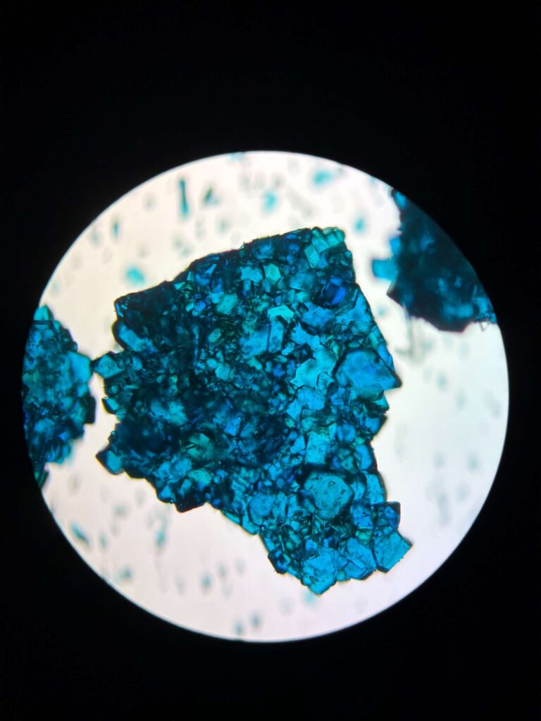
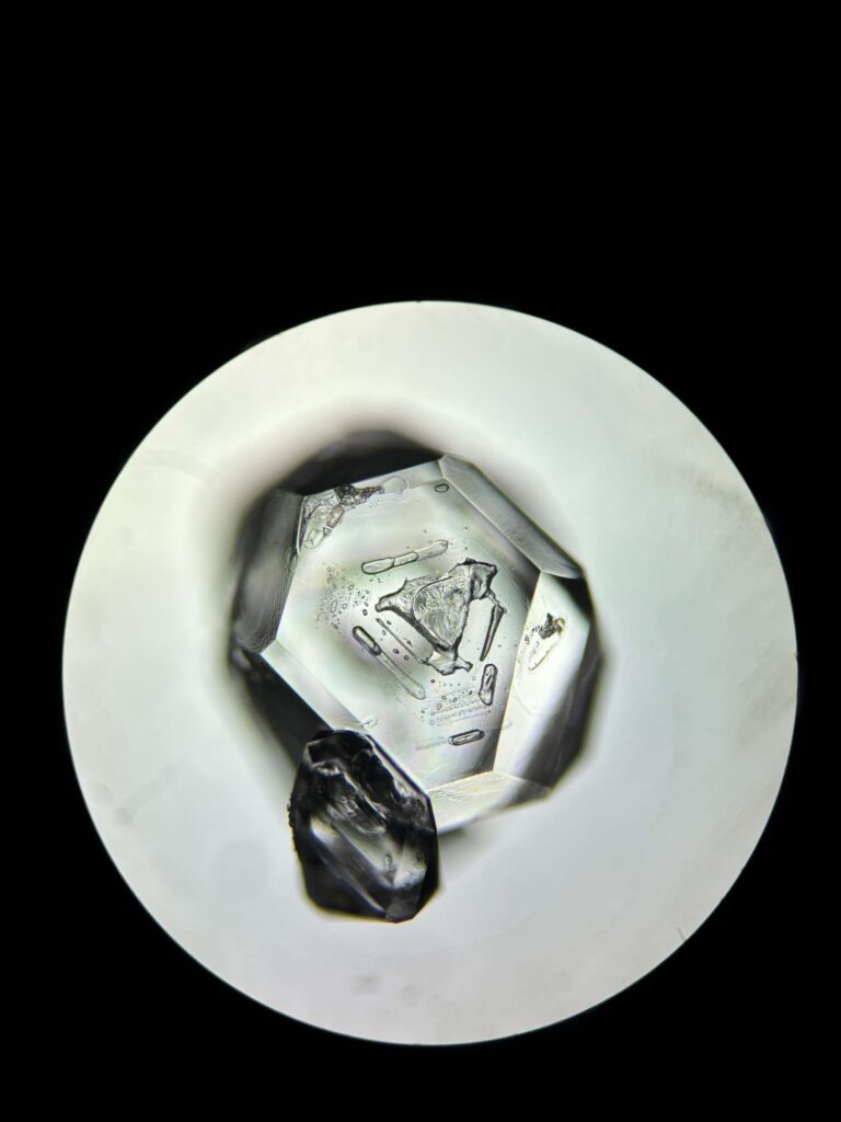
Cell of Alum Alum – Recrystallized potassium alum. Made with Motic/BA210LED Trinocular Microscope (17 mm, 4x/0.10 Zoom) Hidden Suns – Recrystallized potassium alum. Made with Motic/BA210LED Trinocular Microscope (17 mm, 4x/0.10 Zoom) Secret Corridor in the Strawberry – Recrystallized potassium alum. Made with Motic/BA210LED Trinocular Microscope (17 mm, 4x/0.10 Zoom) The Eye of Almighty – Recrystallized potassium alum. Made with Motic/BA210LED Trinocular Microscope (17 mm, 4x/0.10 Zoom) Dancing on the Crater – Recrystallized potassium alum. Made with Motic/BA210LED Trinocular Microscope (17 mm, 4x/0.10 Zoom)
Rokas Vargalis – Institute of Chemistry, Faculty of Geoscience and Chemistry, Vilnius University
I am Rokas Varaglis, a former student of the Faculty of Chemistry and Geosciences. I am currently a chemistry alumnus, and in my free time, one of my many hobbies is imaging various structures, one of these fields is insect photography, using a scanning electron microscope.
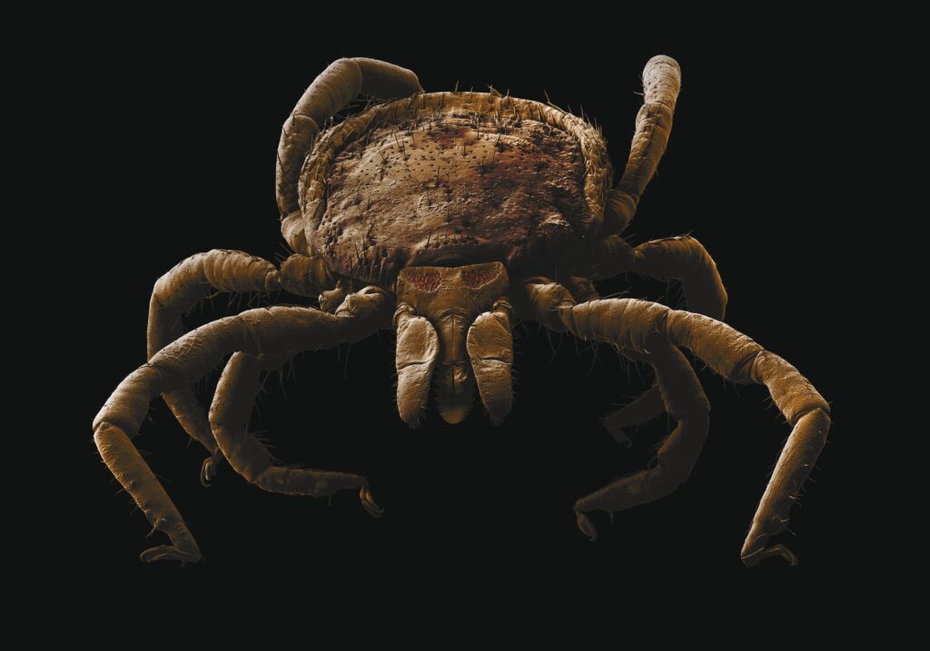
Mite – A mite (Ixodes ricinus) photographed with a scanning electron microscope, processed and colorized using various graphic editing software programs.
Roksana Brzeskot – University of Zielona Góra
I am a third-year Biomedical Engineering student at the University of Zielona Góra. I serve as the vice-chair of the BiomedUZ Scientific Club, where I actively contribute to diverse research projects. My work focuses on pediatric physiotherapy solutions, hydrogel development, and the design of animal prosthetics.
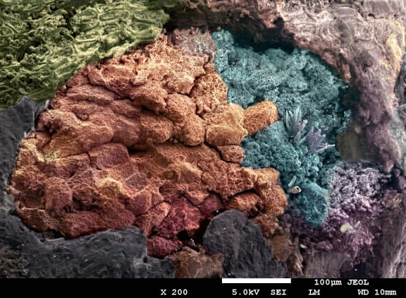
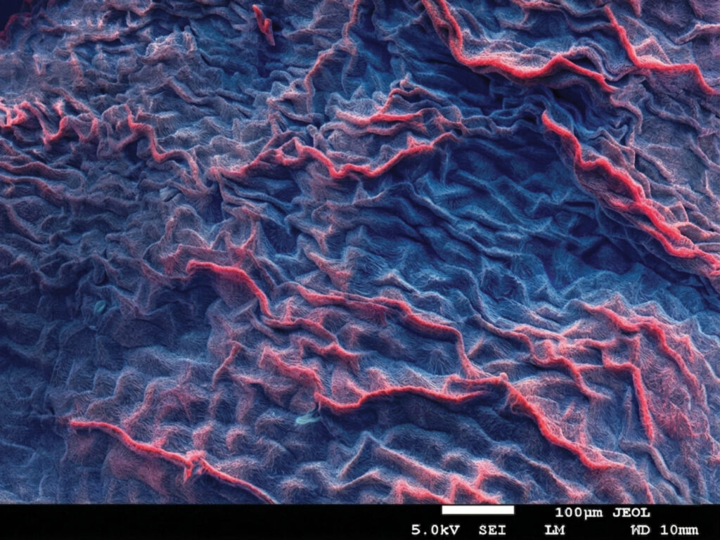
The Coral Reef – This image, captured with a JEOL scanning electron microscope, shows the structure of unroasted buckwheat at 200x magnification. The textures of the endosperm and pericarp create a composition that resembles a coral reef teeming with life. Microscopic Orchard – This SEM image reveals the surface of unroasted buckwheat at high resolution scale bar. Tiny starch granules and plant tissue fragments form patterns that look remarkably like flowers blooming on a tree. Crimson Labyrinth – This microscopic photograph displays the structure of dried chili pepper, captured with a JEOL. Layered edges of the pericarp tissues create an abstract image focused on intense color. Depths of the Unknown – This SEM image shows a fragment of unroasted buckwheat captured with a JEOL. The smooth, flowing contours of the organic matter reveal the natural elasticity and density of the grain’s internal layers.
Samuel Hollý – Cancer Research Institute, Biomedical Research Center, Slovak Academy of Sciences
I am a PhD student at Comenius University in Bratislava with a multidisciplinary background in Optometry, Cell Biology, and Microbiology. I completed a bachelor’s degree in Optometry, a bachelor’s in Experimental and Molecular Biology, and a master’s in Microbiology, all at Masaryk University in Brno. I have research experience with retinal organoids, microRNA biology, and sequencing-based diagnostics, as well as practical clinical optometry experience. My PhD focuses on developing innovative therapeutic approaches for uveal melanoma under the supervision of RNDr. Viera Horváthová Kajabová, PhD. from the Cancer Research Institute (CRI) of the Slovak Academy of Sciences (SAS).
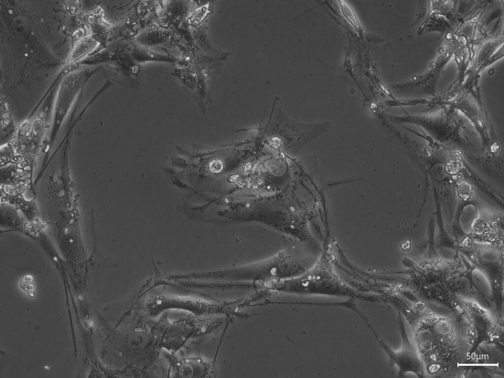
Three Wishes for the Goldfish – Image was acquired using a Mateo FL Digital Fluorescence Microscope (Leica) and shows the pleomorphic MM28 uveal melanoma cell line, derived from a liver metastasis (Uveal Melanoma; Human, Homo sapiens). Cellular Dance — after La Danse by Henri Matisse – Image was acquired using a Mateo FL Digital Fluorescence Microscope (Leica) and shows the pleomorphic MM28 uveal melanoma cell line, derived from a liver metastasis (Uveal Melanoma; Human, Homo sapiens). Raven on a Branch – Image was acquired using a Mateo FL Digital Fluorescence Microscope (Leica) and shows the pleomorphic MM28 uveal melanoma cell line, derived from a liver metastasis (Uveal Melanoma; Human, Homo sapiens). The Heart-Eyed Monster – Image was acquired using a Mateo FL Digital Fluorescence Microscope (Leica) and shows the pleomorphic MM28 uveal melanoma cell line, derived from a liver metastasis (Uveal Melanoma; Human, Homo sapiens).
Sara Niemiec – University of Zielona Góra
Hi, I’m Sara, a 21-year-old third-year Biomedical Engineering student at the University of Zielona Góra. I love dancing, traveling, and spending time with animals. In my free time, I work as a pet sitter.
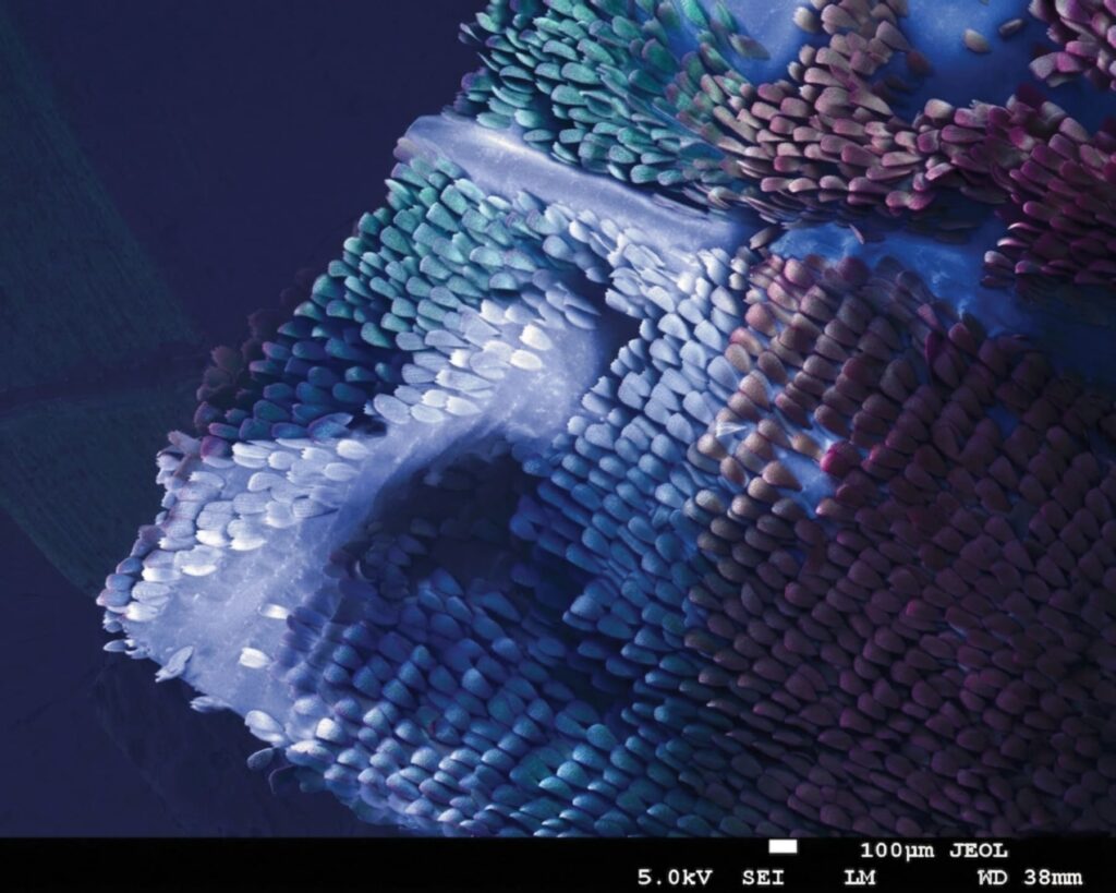
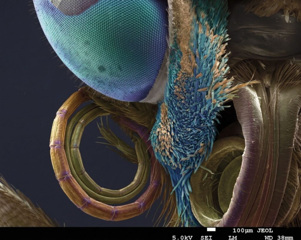
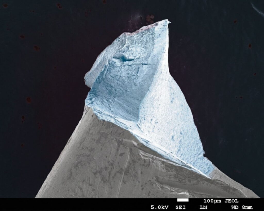
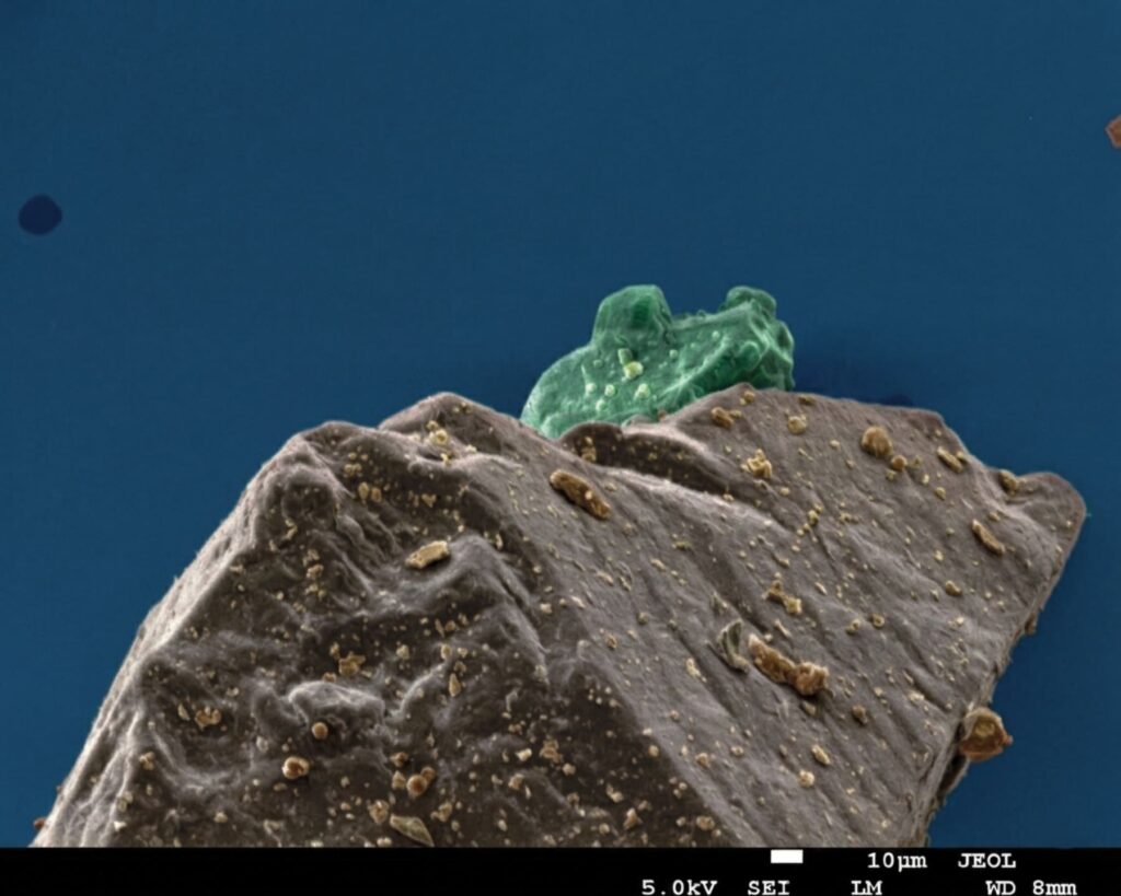
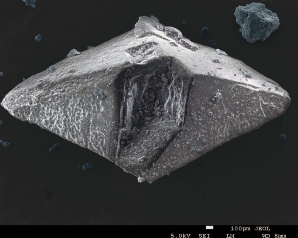
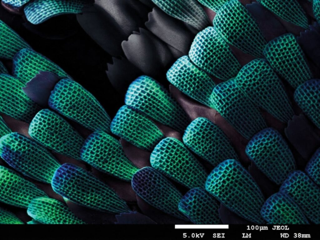
A sequenced wing – This image illustrates the detailed microstructure of a Morpho peleides butterfly wing, captured with a JEOL Scanning Electron Microscope (SEM). The high-resolution scan reveals a dense arrangement of overlapping scales that creates the wing’s signature iridescent shimmer. This natural mosaic resembles a field of colorful sequins, showcasing the complex structural beauty found at a microscopic level. The Butterfly Portrait – This striking close-up presents the intricate anatomy of a Morpho peleides butterfly’s head, captured using a JEOL Scanning Electron Microscope (SEM). The image reveals the remarkable geometric patterns of the compound eye alongside the tightly coiled proboscis, which the insect uses to feed on nectar. By highlighting the vibrant structural scales and delicate sensory hairs, this artwork showcases the complex biological that is invisible to the naked eye. The Microscopic Glacier – This captivating image presents a fractured metal component from a watch, captured using a JEOL Scanning Electron Microscope (SEM). The sharp, jagged edges and icy blue tones of the metallic surface create a striking resemblance to a massive glacier. By revealing the hidden topography of everyday objects, this scan transforms a tiny fragment of machinery into a monumental landscape of industrial beauty. The Frog Hidden in a Tablet – This intriguing image showcases a particle of the Biolevox dietary supplement, captured using a JEOL Scanning Electron Microscope (SEM). The unique formation of the mineral structures creates a whimsical scene that strongly resembles a small green frog perched atop a rugged mountain peak. By enlarging these microscopic components, the scan reveals a surprising and artistic landscape hidden within a tablet. The Spacecraft Within the Tablet – This extraordinary image presents a microscopic particle from the Biolevox supplement, visualized through a JEOL Scanning Electron Microscope (SEM). The sharp, angular geometry and central indentation of the mineral structure create a striking resemblance to a futuristic spacecraft drifting through the void of space. By exploring these tiny components at high magnification, the scan reveals a cosmic landscape hidden within a tablet.
Stefania Wolff – Faculty of Applied Physics and Mathematics, Gdańsk University of Technology and Centre of Plasma and Laser Engineering, Institute of Fluid-Flow Machinery, Polish Academy of Sciences
I am a PhD student at Gdańsk University of Technology and work at the Institute of Fluid-Flow Machinery, Polish Academy of Sciences. My research focuses on titania nanotube based electrodes and their applications in photoelectrochemistry, including oxygen and hydrogen generation.
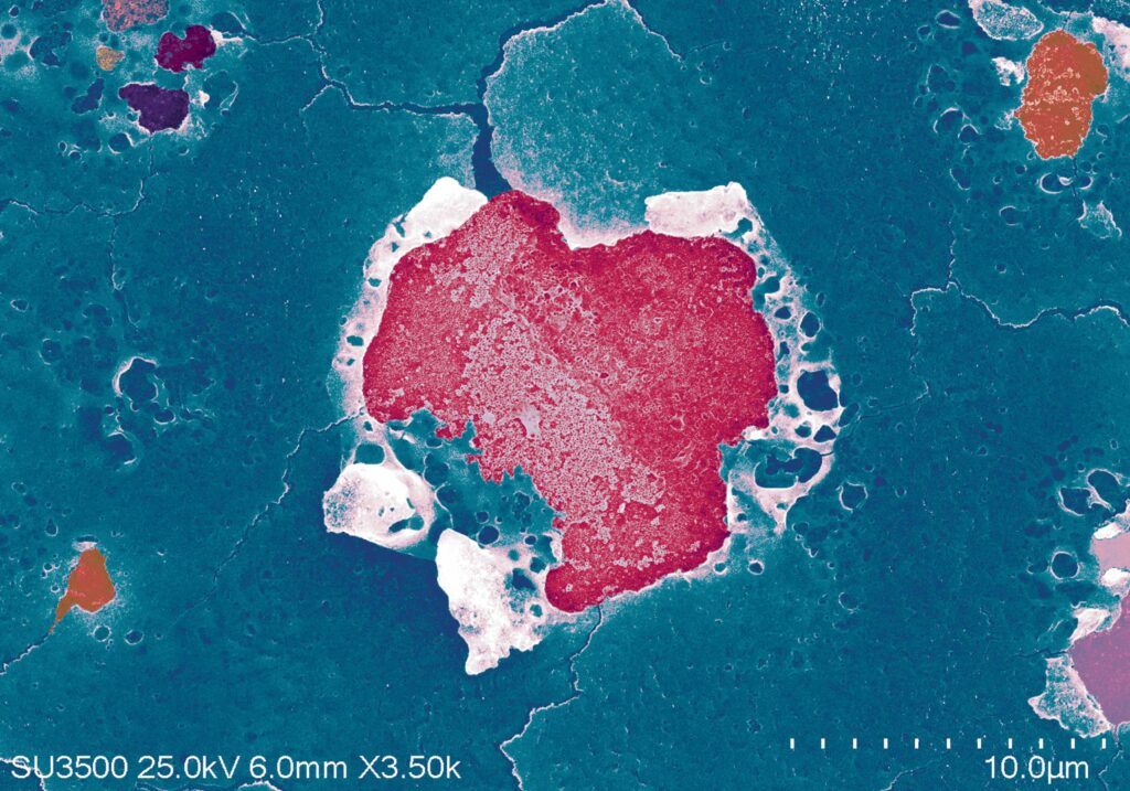
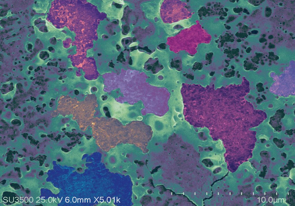
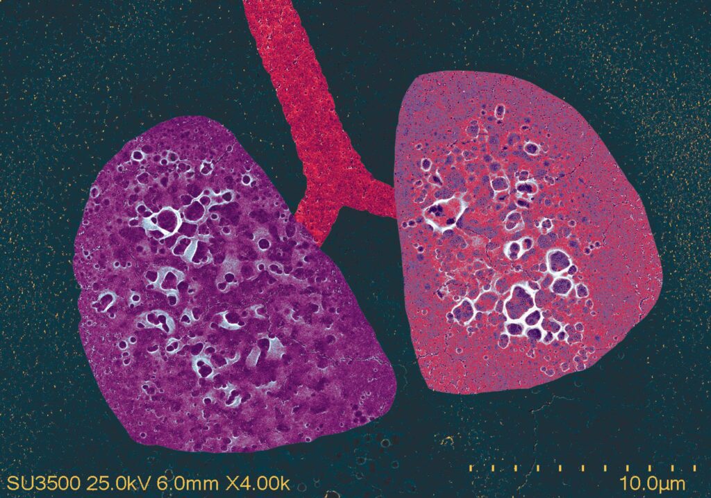
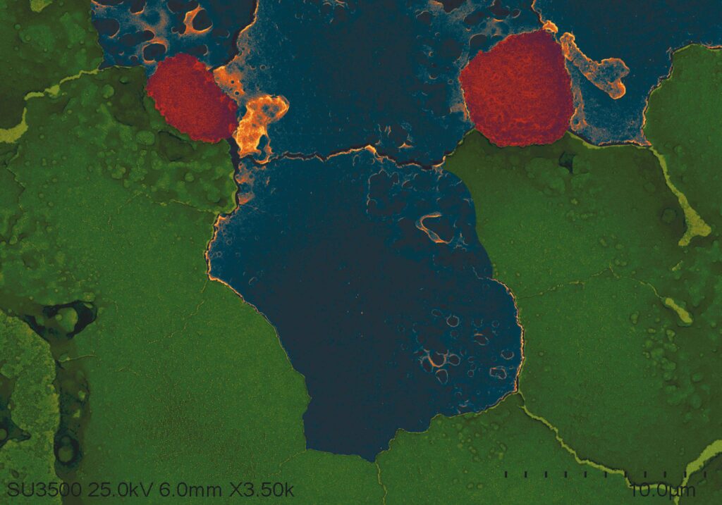
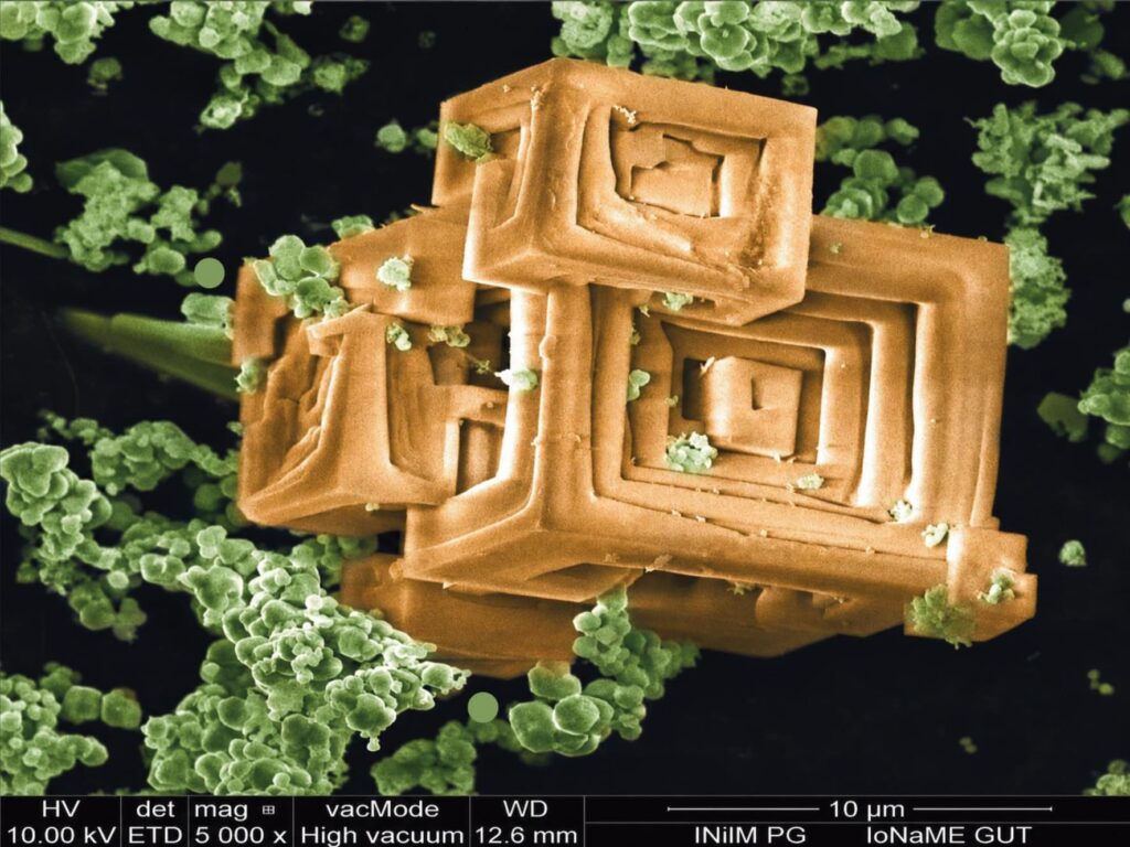
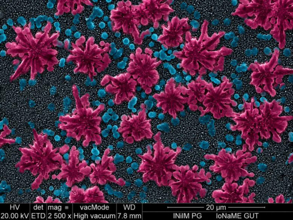
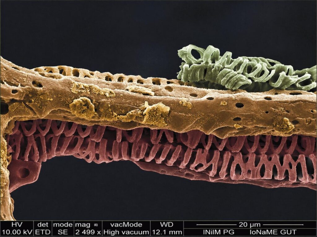
Matryoshka – Colorized scanning electron microscope (SEM, FEI Quanta FEG 250, FEI Company) image showing residual salt formed during the synthesis of BaTiO3. Nanojungle – Colorized scanning electron microscope (SEM, FEI Quanta FEG 250, FEI Company) image showing the cross-section of titanium dioxide nanotubes modified with palladium. Nanoflowers – Colorized scanning electron microscope (SEM, FEI Quanta FEG 250, FEI Company) image showing the surface of titanium nanotubes modified with tungsten sulfide. Chameleon – Colorized scanning electron microscope (SEM, FEI Quanta FEG 250, FEI Company) image of a Ficus religiosa leaf. Bubbles – Colorized scanning electron microscope (SEM, FEI Quanta FEG 250, FEI Company) image showing the surface of titanium nanotubes modified with tungsten sulfide.
Teun De Voeght – University of Antwerp
I work for the Faculty Pharmaceutical, Biomedical and Veterinary Sciences as a digital content creator, responsible for the content on our various social media channels. I’m the one who’s always walking around with a camera, sticking a microphone under people’s noses. I have a background in journalism and worked as a freelancer for many years before joining the University of Antwerp. I studied International Politics and Journalism and have a passion for the natural world.
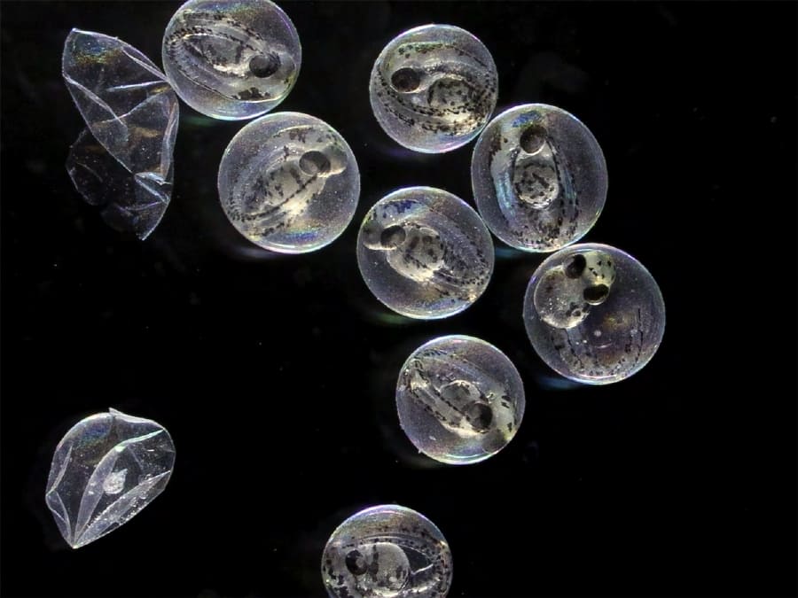
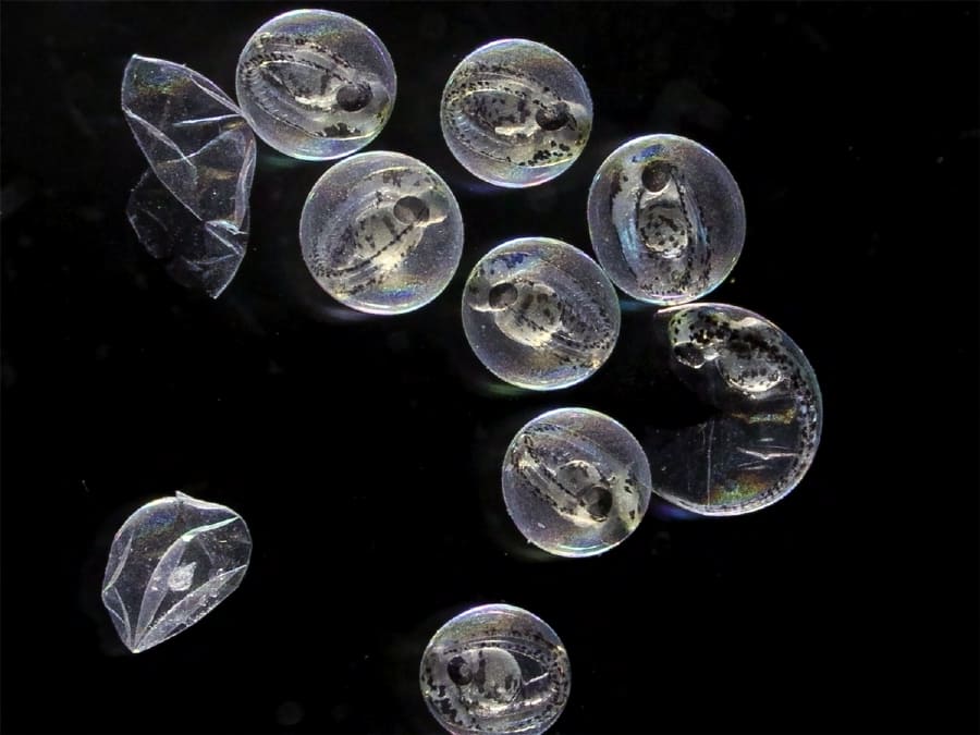
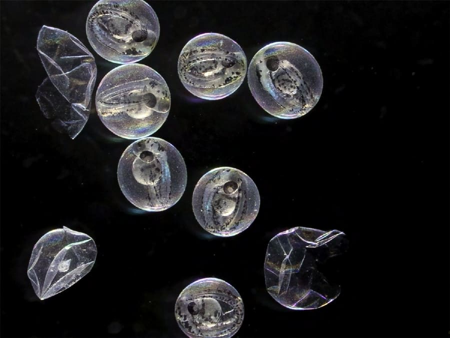
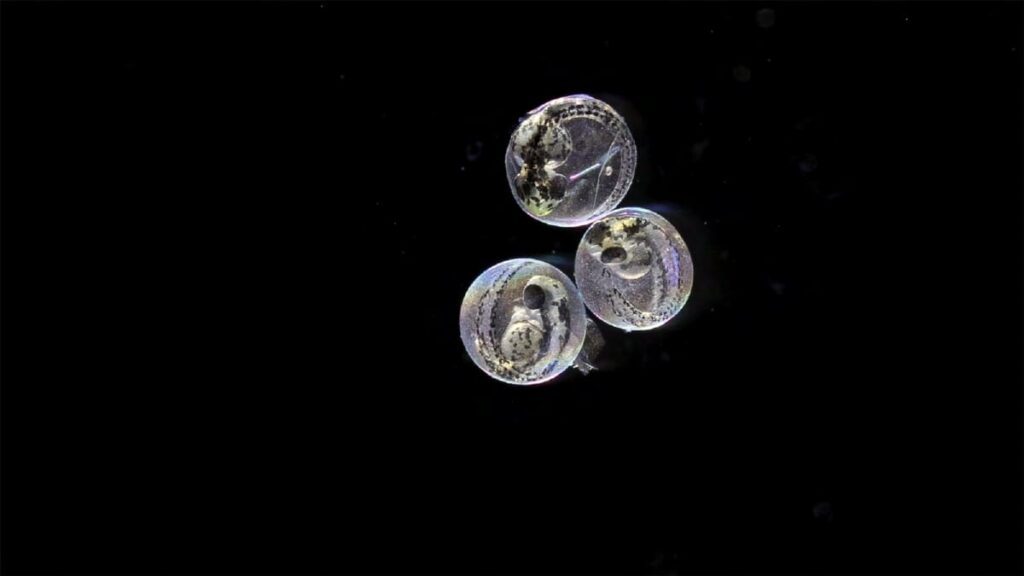
The Birth of a Zebrafish IThe Birth of a Zebrafish IIThe Birth of a Zebrafish IIIThe Birth of a Zebrafish IV
Tola Granowska – Wrocław University of Environmental and Life Sciences
Biology first year student 🙂
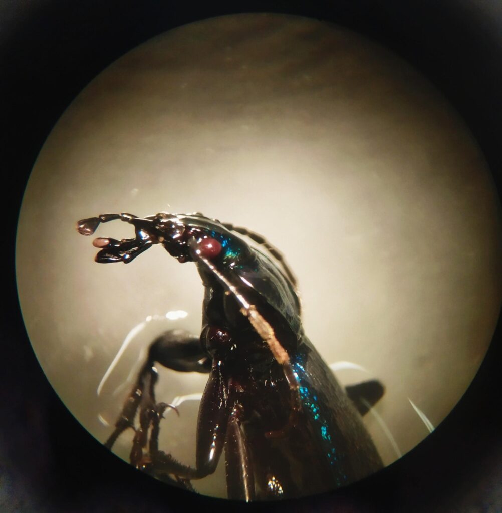
Prince of Darkness – Side profile of Carabus insect. Biologist’s moon – Arm-palisade parenchyma cells. Mouse steak – Mouse liver tissue. What goes on underneath – Mouse skin tissue. Crystal desert – Salt crystals.
Wiktoria Buczyńska – University of Zielona Góra
I am a third-year Biomedical Engineering student at the University of Zielona Góra and the president of the BiomedUZ student research club. I am deeply passionate about science, 3D design, and laboratory work, where I can combine curiosity with hands-on experimentation and problem-solving.
Microscopic Coral Reef – This false-color scanning electron micrograph (SEM), captured using a JEOL Scanning Electron Microscope, reveals the intricate surface topography of an orchid flower at high magnification. The image highlights complex microstructures resembling an underwater coral reef landscape. Micro Bloom – Using a JEOL Scanning Electron Microscope, this image reveals the delicate surface of a tiny flower. The textures resemble a blooming miniature landscape. Micro Bloom – Using a JEOL Scanning Electron Microscope, this image reveals the delicate surface of a tiny flower. The textures resemble a blooming miniature landscape. Butterfly Mosaic – Taken with a JEOL Scanning Electron Microscope, the false-color SEM image of a Papilio palinurus butterfly wing displays tightly packed scales forming a natural mosaic. Ocean of the Orchid – This false-color scanning electron micrograph (SEM), captured using a JEOL Scanning Electron Microscope, presents the velvety surface of an orchid petal in false color, with patterns that look like waves and underwater formations.
Wiktoria Michalik – Institute of Materials Engineering, Faculty of Science and Technology, University of Silesia in Katowice
I am a PhD student at the International Environmental Doctoral School at the University of Silesia in Katowice. My research focuses on metallic materials, with particular emphasis on composites for biomedical applications. I am a fan of science fiction films, and in my free time I enjoy playing video games and crocheting plush toys.
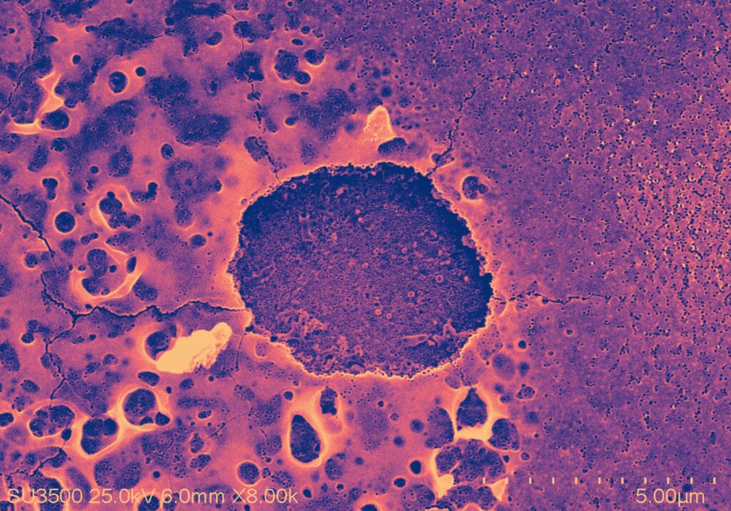
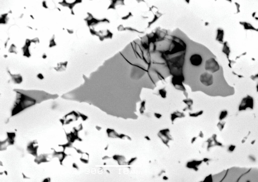
Radioactive Fish – Microstructure of a titanium matrix composite reinforced with TiMo particles, obtained using a scanning electron microscope (SEM JEOL JSM–6480). Backscattered electrons (BSE) were used for imaging, and the resulting contrast between the regions reveals what at first glance resembles a fish with three pairs of eyes. Crying Face – Microstructure of a titanium matrix composite reinforced with TiMo particles, obtained using a scanning electron microscope (SEM JEOL JSM–6480). The porosity present in the microstructure formed in an unusual arrangement, revealing a smiling yet crying face. Imaginary Coastline – An image of a stone obtained using a Keyence VHX-X1 digital microscope. The resulting image resembles a bird’s-eye view of a coastline, while the colorful spots appear similar to beachgoers. Something Resembling a “Lava Lamp” – Microstructure of a gradient titanium matrix composite reinforced with TiMo particles obtained using a Keyence VHX-X1 digital microscope. The boundary between the two layers resembles a “lava lamp”. Meteorite-like Porosity – Microstructure of a titanium matrix composite reinforced with TiMo particles, obtained using a scanning electron microscope (SEM JEOL JSM–6480). The nearly circular pore visible in the image resembles a meteorite moving at high speed toward the upper left corner.
Wioletta Lech – Mossakowski Medical Research Institute, Polish Academy of Sciences
I work at the Department of Stem Cell Bioengineering, Mossakowski Medical Research Institute, Polish Academy of Sciences in Warsaw. I conduct research in the field of stem cell biology, creation of a three-dimensional microenvironment in vitro that mimics in vivo conditions, isolation of therapeutically competent stem cells, and evaluation of their neuroprotective, pro-regenerative, and immunomodulatory properties in preclinical translational studies. In my research, I mainly focus on the use of stem cells in the treatment of central nervous system disorders and the implementation of advanced therapy medicinal products for clinical use.
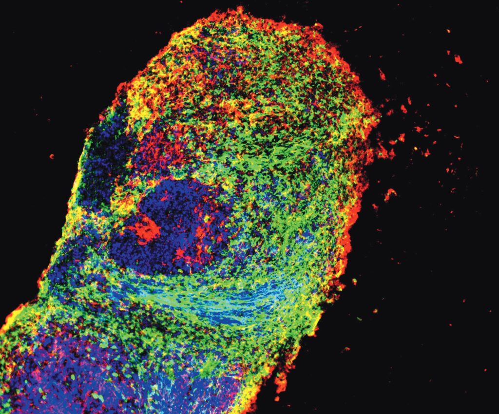
The Unknown Lifeform – Immunocytochemical characterization of 40-day human Dorsal Forebrain Organoid for neural markers – Nestin (green) and Doublecortin (red). Cell nuclei were labelled with Hoechst 33342 (blue). ZEISS Axiovert 5 digital All-in-One Cell Imaging System was used for image acquisition.
Wojciech Piotrowski – Institute of Advanced Materials, Faculty of Chemistry, Wrocław University of Science and Technology
I received my PhD from ILTSR Polish Academy of Sciences in 2024 and currently work at Wrocław Tech. My research centers on near-infrared luminescence in nanoparticles for imaging and sensing applications.
Nanoscience loves back – Scanning electron microscope (SEM) image of NaYF4 nanoparticles. The image was acquired by Dr Wojciech Gil (University of Wroclaw) using a Thermo Scientific SCIOS 2 microscope.
Zofia Mazurkiewicz – Institute of Low Temperature and Structure Research, Polish Academy of Sciences
Chemist and radiation chemistry laboratory supervisor. She is responsible for the synthesis and preparation of samples for microscopic measurements.
Żaneta Walendziak – Warsaw University of Technology
I am a master’s student in Materials Engineering at the Warsaw University of Technology. In my free time, I work on algae cultivation.
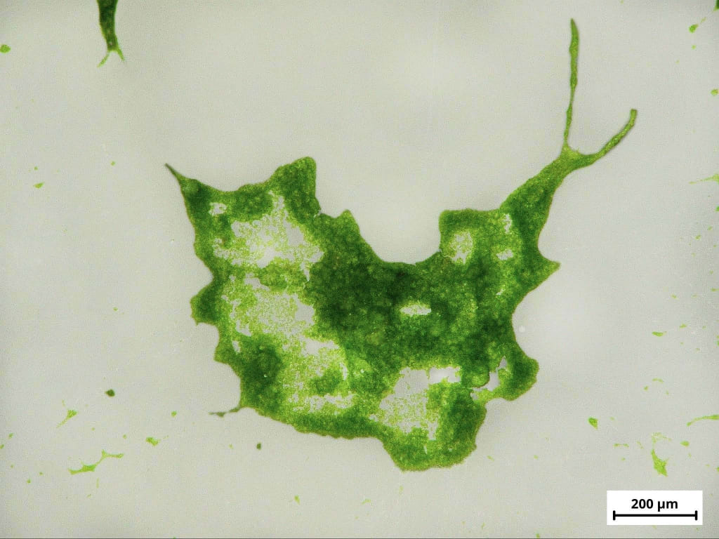
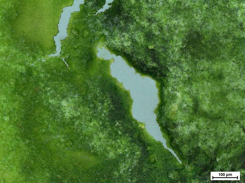
Algae snail – The photo shows a snail-shaped algal biofilm (Chlorella vulgaris) on a polymer sample after 7 days of incubation. The photo was taken with Keyence VHX970FN Digital Microscope. A landscape of algae – The photo shows an algal biofilm (Chlorella vulgaris) resembling mountains and a lake on a polymer sample after 7 days of incubation. The photo was taken with Keyence VHX-7000 Digital Light Microscope, 300x magnification.